Uncategorized files
Jump to navigation
Jump to search
Showing below up to 50 results in range #51 to #100.
-
 Blue circle for diabetes.svg.png 240 × 240; 8 KB
-
 Bluthirnschranke nach Infarkt nativ und KM.png 1,590 × 984; 549 KB
-
 BroadTachydiff.png 1,200 × 949; 90 KB
-
 Brugada.jpg 126 × 160; 13 KB
-
 Brugada.png 800 × 973; 29 KB
-
 Brugada ecg characteristics.png 1,000 × 750; 39 KB
-
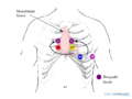 Brugada lead placement.png 800 × 600; 84 KB
-
 Brugada syndrome type1 example1.png 3,300 × 1,949; 370 KB
-
 Brugada syndrome type1 example2.png 3,307 × 1,949; 348 KB
-
 Brugada syndrome type1 example3.png 3,300 × 1,949; 63 KB
-
 Brugada syndrome type1 example4.png 3,300 × 1,950; 364 KB
-
 Brugada syndrome type1 example5.png 3,300 × 1,950; 364 KB
-
 Brugada syndrome type1 example6.jpg 3,249 × 2,131; 970 KB
-
 Brugada syndrome type2 example1.png 3,300 × 1,950; 363 KB
-
 Brugada syndrome type2 example2.jpg 3,485 × 1,891; 677 KB
-
 CPVT.jpg 566 × 472; 34 KB
-
 CPVT.svg 575 × 473; 467 KB
-
 CRT device.svg 1,035 × 615; 443 KB
-
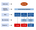
 CRT flowchart.svg 1,200 × 1,358; 55 KB
-
 Cardiac injury myocarditis.png 712 × 571; 108 KB
-
 Cardiac injury myocarditis.svg 721 × 577; 83 KB
-
 Cardiac markers.png 839 × 590; 56 KB
-
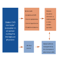 Cardiologist menopause physician.svg 1,200 × 1,200; 22 KB
-
 Cardionetworks.png 437 × 39; 6 KB
-
 Causes of myocarditis.png 1,000 × 1,024; 64 KB
-
 Causes of myocarditis.svg 774 × 960; 71 KB
-
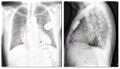 ChestXray.jpg 595 × 342; 46 KB
-
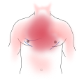 Chest pain areas.svg 1,200 × 1,200; 20 KB
-
 Chest pain to NSTEMI STEMI.svg 1,200 × 1,000; 28 KB
-
 Chest pain to NSTEMI STEMI v2.svg 940 × 860; 340 KB
-
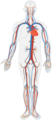
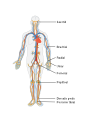 Circulatory System.svg 931 × 1,313; 368 KB
-
 Circulatory System no tags.svg 259 × 599; 104 KB
-
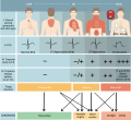
 Clinical scenarios.jpg 985 × 1,015; 179 KB
-
 Clinical scenarios.svg 966 × 897; 65 KB
-
 Conductionsystem.svg 1,000 × 750; 201 KB
-
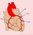 Coronary anatomy.png 800 × 850; 142 KB
-
 DPP6ECG.jpg 567 × 443; 218 KB
-
 Ddd paced 12lead.jpg 800 × 508; 73 KB
-
 Debakey.svg 512 × 360; 33 KB
-
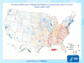 Diabetes County level estimates 2004-2009.gif 800 × 600; 363 KB
-
 Diabetes mellitus world map - DALY - WHO2004.svg 800 × 353; 78 KB
-
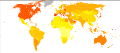 Diabetes world map - 2000.png 940 × 415; 1.45 MB
-
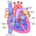 Diagram of the human heart (valves improved).svg 650 × 650; 27 KB
-
 Differences in coronary artery disease by gender.svg 1,200 × 1,200; 221 KB
-
 Dissections.svg 512 × 382; 15 KB
-
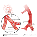
 Distalembolization.svg 1,320 × 764; 252 KB
-
 ECGT.jpg 686 × 212; 57 KB
-
 ECG SitusInversus ElektrodesPlacedLeft.JPG 1,697 × 1,261; 1.24 MB
-
 ECG SitusInversus ElektrodesPlacedRight.JPG 1,951 × 1,440; 1.61 MB
-
 Endo dysfunction.png 1,180 × 1,296; 958 KB


.svg)


