File:Gray1216 modern locations.svg
Jump to navigation
Jump to search

Size of this PNG preview of this SVG file: 633 × 600 pixels.
Original file (SVG file, nominally 645 × 611 pixels, file size: 25 KB)
| Description |
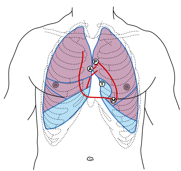
Front of thorax, showing surface relations of bones, lungs (purple), pleura (blue), and heart (red outline). Heart valves are labeled (Mitral (Bicuspid), Tricuspid, Aortic, Pulmonary). Figure 1216 from Gray's Anatomy. It has been updated to reflect modern thinking on where heart sounds are best heard. |
|---|---|
| Source | |
| Date |
2010-02-26 03:58 (UTC) |
| Author |
|
| Permission |
File history
Click on a date/time to view the file as it appeared at that time.
| Date/Time | Thumbnail | Dimensions | User | Comment | |
|---|---|---|---|---|---|
| current | 09:15, 5 December 2012 | | 645 × 611 (25 KB) | Drj (talk | contribs) | {{Information |Description=Front of thorax, showing surface relations of bones, lungs (purple), pleura (blue), and heart (red outline). Heart valves are labeled ('''M'''itral ('''B'''icuspid), '''T'''ricuspid, '''A'''ortic, '''P'''ulmonary). Figure 121... |
You cannot overwrite this file.
File usage
The following page uses this file: