File:Figure 9.jpg
Jump to navigation
Jump to search
Size of this preview: 742 × 599 pixels. Other resolutions: 2,536 × 2,048 pixels | 4,606 × 3,720 pixels.
Original file (4,606 × 3,720 pixels, file size: 1.19 MB, MIME type: image/jpeg)
| Description |
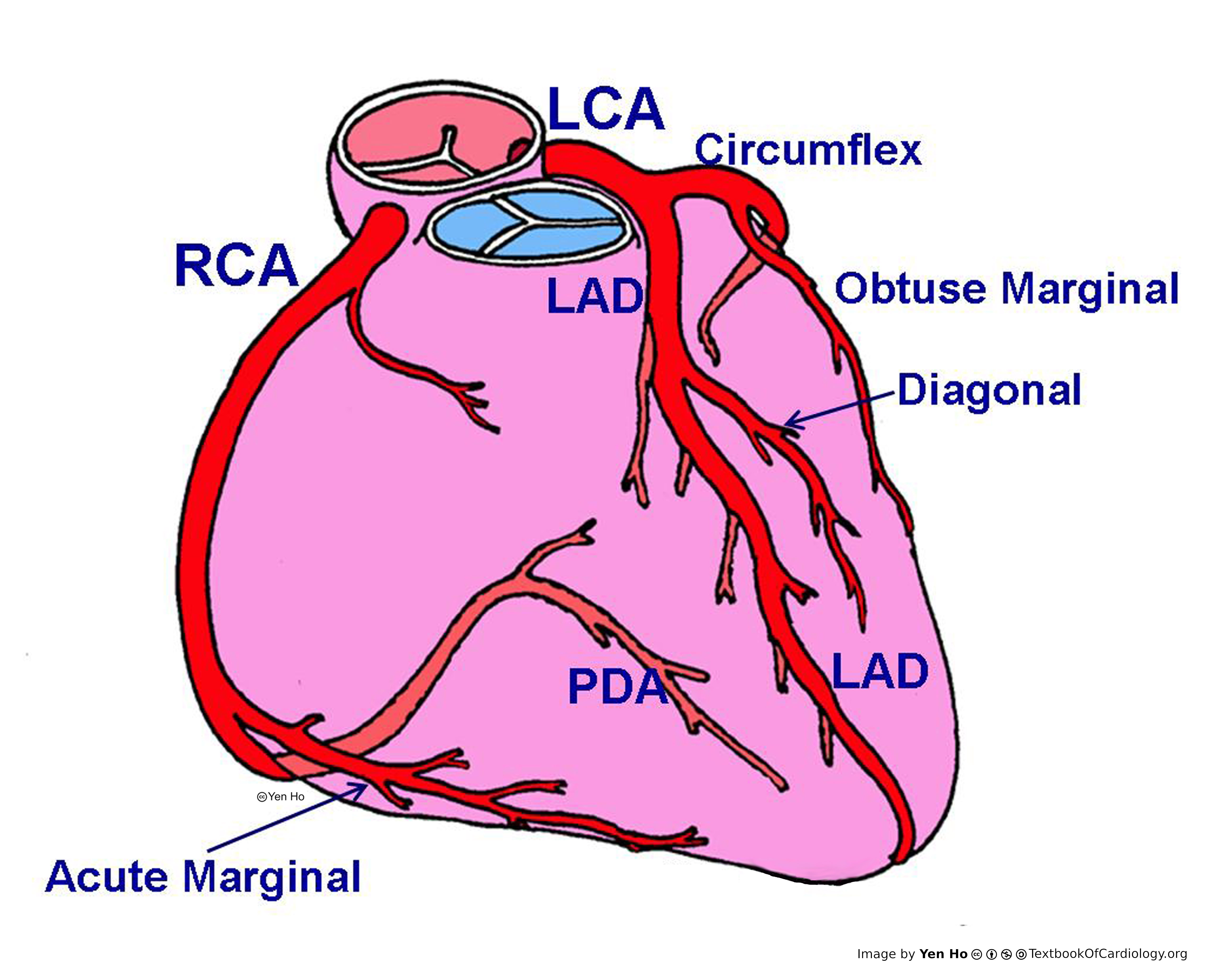
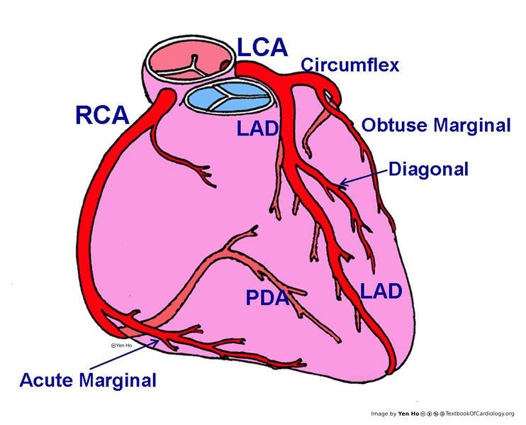
Diagram showing the right (RCA) and left (LCA) coronary arteries and their main ventricular branches. The left anterior descending (LAD) and posterior descending (PDA) coronary arteries mark the anterior and posterior margins of the ventricular septum. |
|---|---|
| Source |
provided by S. Yen Ho, PhD FRCPath FESC FHEA, Royal Brompton Hospital, UK |
| Date |
2012 |
| Author |
S. Yen Ho, PhD FRCPath FESC FHEA, Royal Brompton Hospital, UK |
| Permission |
File history
Click on a date/time to view the file as it appeared at that time.
| Date/Time | Thumbnail | Dimensions | User | Comment | |
|---|---|---|---|---|---|
| current | 11:35, 18 May 2012 | | 4,606 × 3,720 (1.19 MB) | NiloferT (talk | contribs) | |
| 11:18, 18 May 2012 | No thumbnail | (1.19 MB) | NiloferT (talk | contribs) |
You cannot overwrite this file.
File usage
There are no pages that use this file.