File:Figure 9. Schematic drawing of the anatomy prenatal and postnatal.png

Original file (1,166 × 868 pixels, file size: 1.24 MB, MIME type: image/png)
| Description |
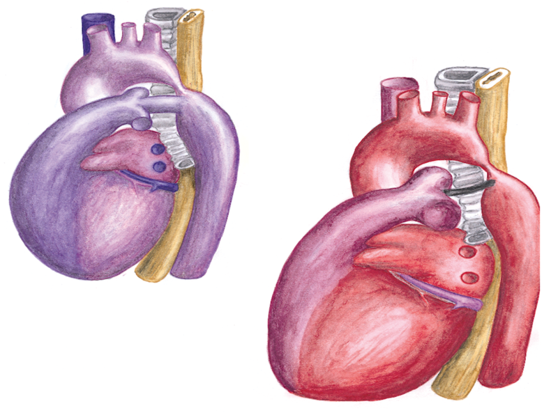
Figure 9. Schematic drawing of the anatomy prenatal (left) and postnatal (right) in coarctation of the aorta. In the normal situation (without coarctation) only 10 percent of the fetal cardiac output flows through the descending aorta. Therefore there are no hemodynamic consequences prenatal of coarctation of the aorta. In the postnatal situation, after closure of the ductus arteriosus, around 75% of cardiac output needs to pass the coarctation, leading to obstruction. |
|---|---|
| Source |
illustration by dr. J.P.M. Hamer |
| Date | |
| Author |
illustration by dr. J.P.M. Hamer |
| Permission |
with permission |
File history
Click on a date/time to view the file as it appeared at that time.
| Date/Time | Thumbnail | Dimensions | User | Comment | |
|---|---|---|---|---|---|
| current | 16:10, 1 February 2012 | | 1,166 × 868 (1.24 MB) | Nja (talk | contribs) | {{Information |Description=Figure 9. Schematic drawing of the anatomy prenatal (left) and postnatal (right) in coarctation of the aorta. In the normal situation (without coarctation) only 10 percent of the fetal cardiac output flows through the descending |
You cannot overwrite this file.
File usage
The following page uses this file: