File:Figure 6.jpg
Original file (1,772 × 3,104 pixels, file size: 601 KB, MIME type: image/jpeg)
| Description |
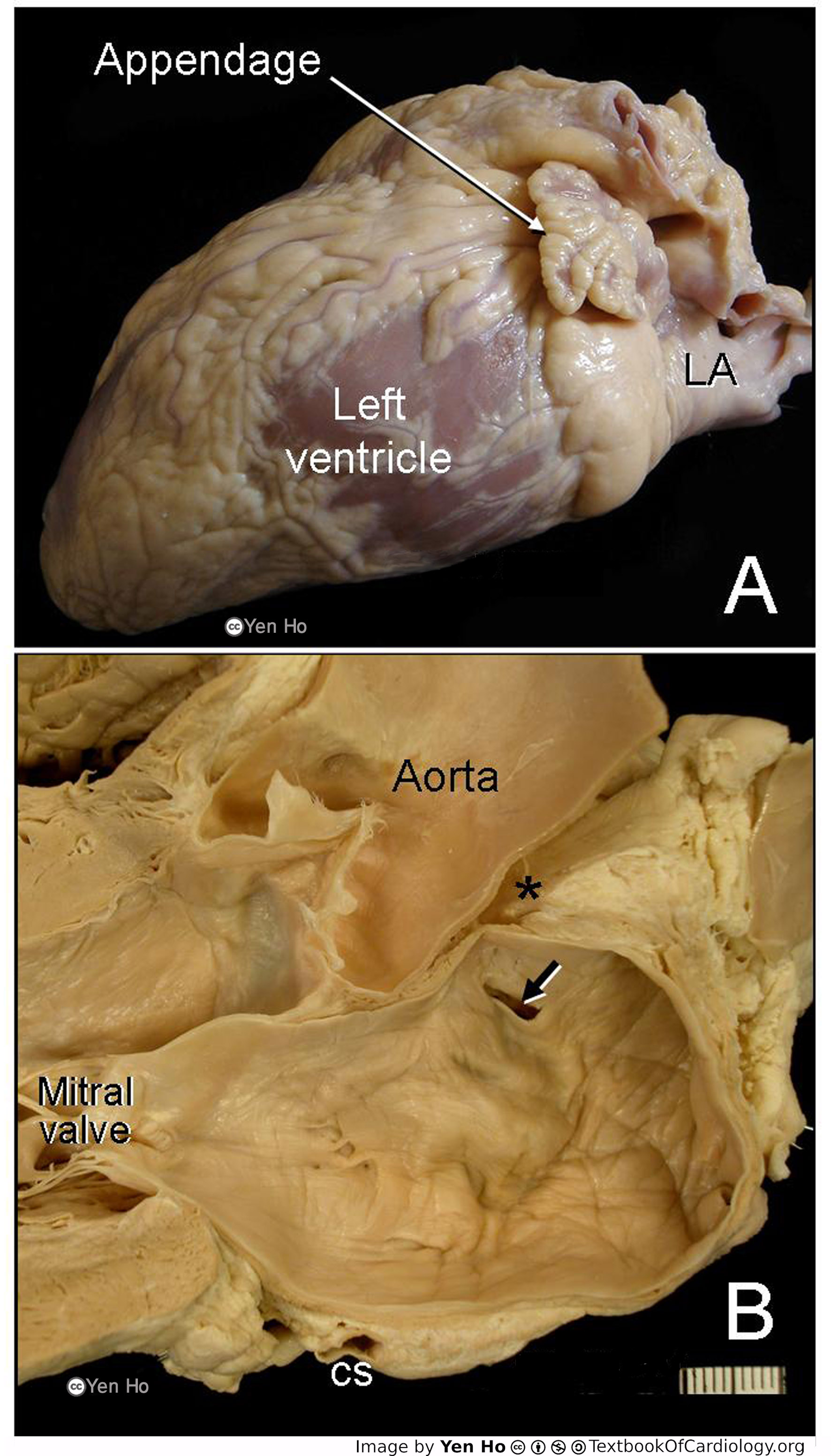
A. This view from the left-lateral aspect shows the finger-like left atrial appendage with the left atrium situated posteriorly. The left ventricle tapers to a rounded apex.
|
|---|---|
| Source |
provided by S. Yen Ho, PhD FRCPath FESC FHEA, Royal Brompton Hospital, UK |
| Date |
2012 |
| Author |
S. Yen Ho, PhD FRCPath FESC FHEA, Royal Brompton Hospital, UK |
| Permission |
File history
Click on a date/time to view the file as it appeared at that time.
| Date/Time | Thumbnail | Dimensions | User | Comment | |
|---|---|---|---|---|---|
| current | 10:50, 18 May 2012 | | 1,772 × 3,104 (601 KB) | NiloferT (talk | contribs) |
You cannot overwrite this file.
File usage
The following page uses this file: