File:Figure 5.jpg
Original file (3,543 × 2,104 pixels, file size: 815 KB, MIME type: image/jpeg)
| Description |
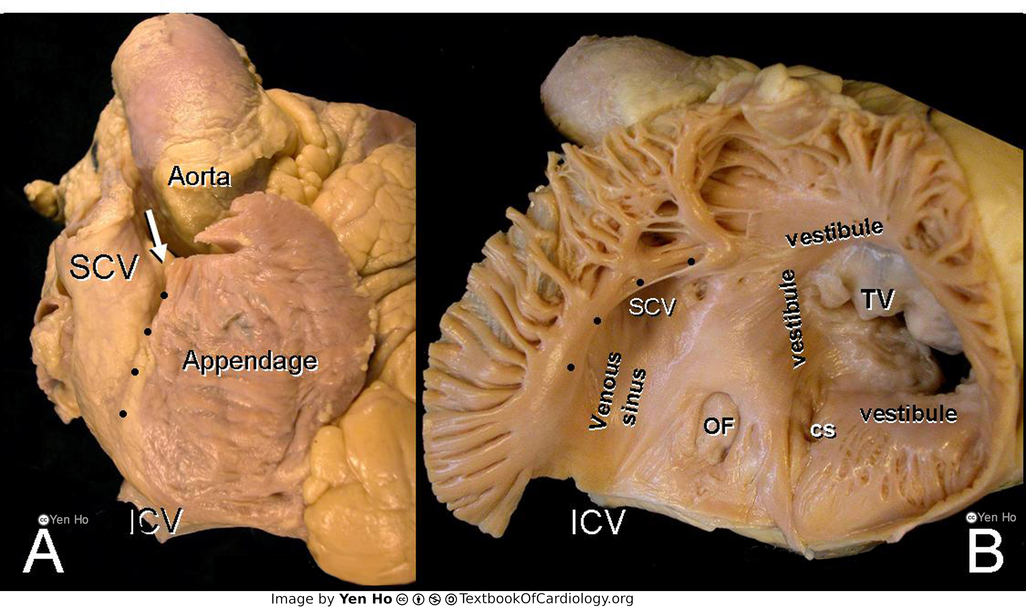
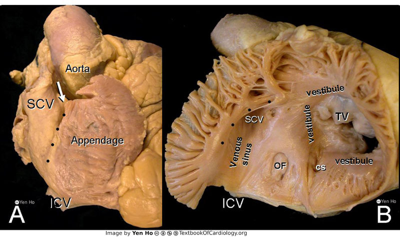
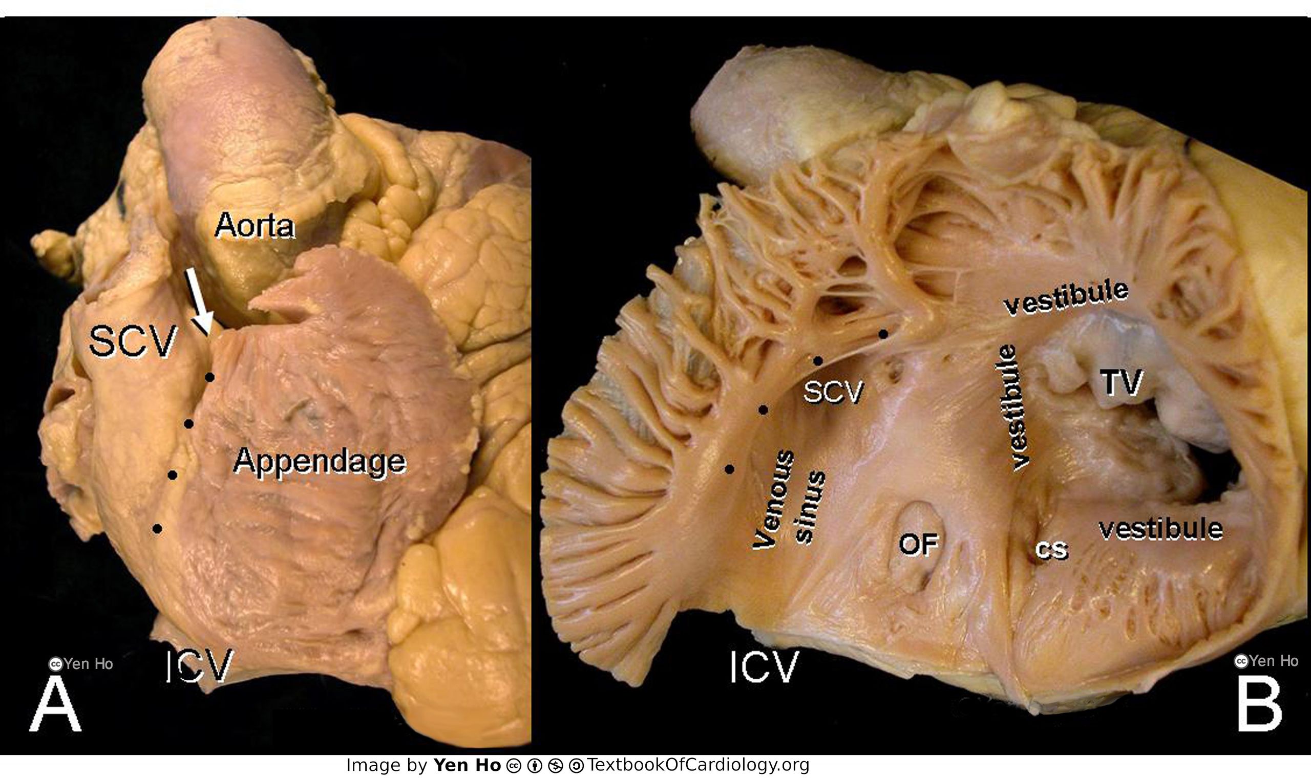
A. This right lateral view shows the right atrium dominated by its large, triangluar shaped appendage. The dots mark the terminal groove. The arrow indicates the crest of the appendage.
|
|---|---|
| Source |
provided by S. Yen Ho, PhD FRCPath FESC FHEA, Royal Brompton Hospital, UK |
| Date |
2012 |
| Author |
S. Yen Ho, PhD FRCPath FESC FHEA, Royal Brompton Hospital, UK |
| Permission |
File history
Click on a date/time to view the file as it appeared at that time.
| Date/Time | Thumbnail | Dimensions | User | Comment | |
|---|---|---|---|---|---|
| current | 12:43, 20 May 2012 | | 3,543 × 2,104 (815 KB) | NiloferT (talk | contribs) |
You cannot overwrite this file.
File usage
The following page uses this file: