File:Figure 4.jpg
Jump to navigation
Jump to search
Size of this preview: 398 × 599 pixels. Other resolution: 2,126 × 3,199 pixels.
Original file (2,126 × 3,199 pixels, file size: 566 KB, MIME type: image/jpeg)
| Description |
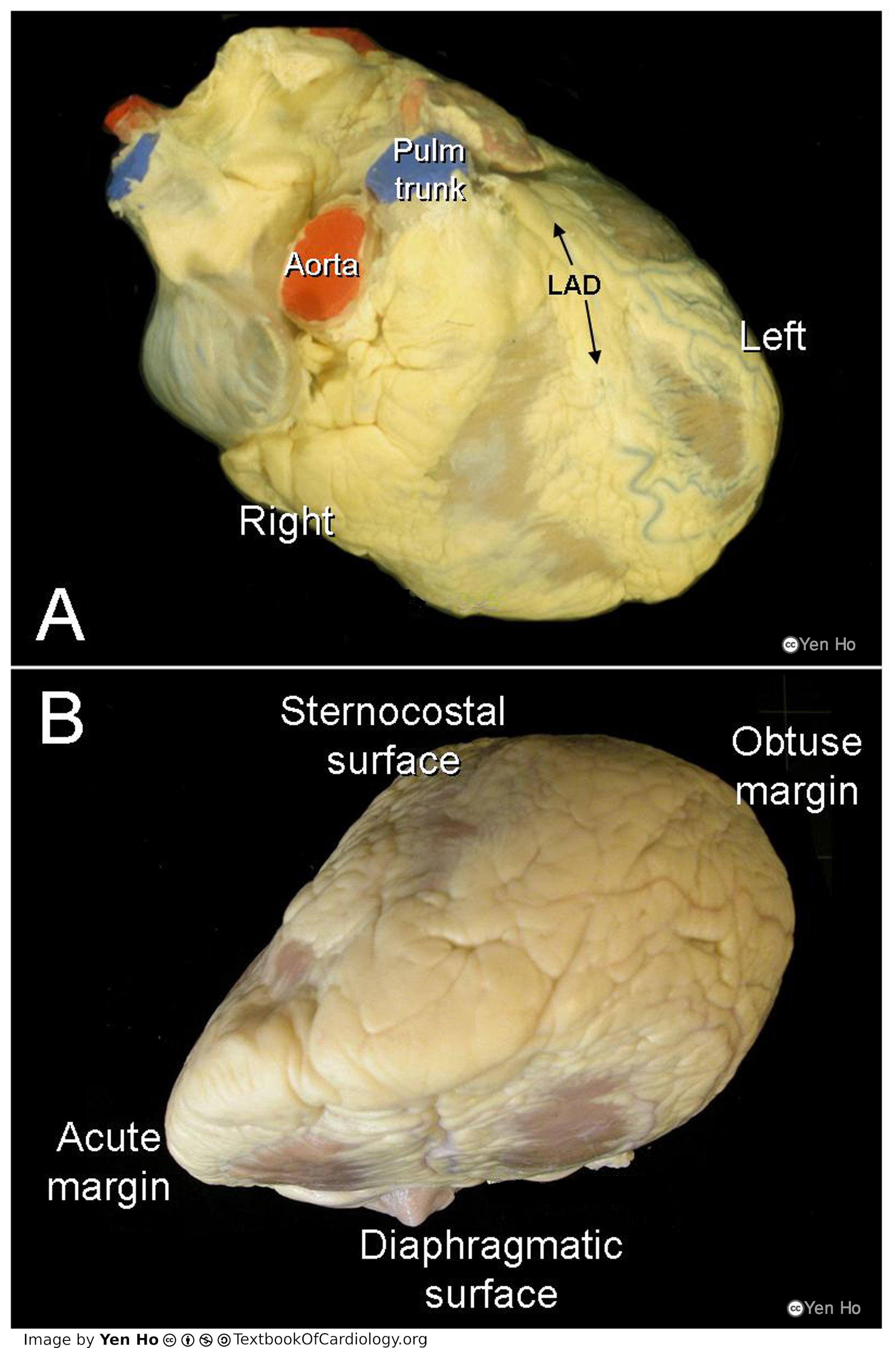
'A. This frontal view shows the right and left surfaces of the heart. The left anterior descending coronary artery buried in epicardial fat marks the plane of the ventricular septum.
|
|---|---|
| Source |
provided by S. Yen Ho, PhD FRCPath FESC FHEA, Royal Brompton Hospital, UK |
| Date |
2012 |
| Author |
S. Yen Ho, PhD FRCPath FESC FHEA, Royal Brompton Hospital, UK |
| Permission |
File history
Click on a date/time to view the file as it appeared at that time.
| Date/Time | Thumbnail | Dimensions | User | Comment | |
|---|---|---|---|---|---|
| current | 10:46, 18 May 2012 | | 2,126 × 3,199 (566 KB) | NiloferT (talk | contribs) |
You cannot overwrite this file.
File usage
The following page uses this file: