File:Figure 21. Schematic drawing showing Ebstein’s anomaly of the tricuspid valve.png
Jump to navigation
Jump to search
Size of this preview: 779 × 600 pixels. Other resolution: 1,462 × 1,126 pixels.
Original file (1,462 × 1,126 pixels, file size: 1.44 MB, MIME type: image/png)
| Description |
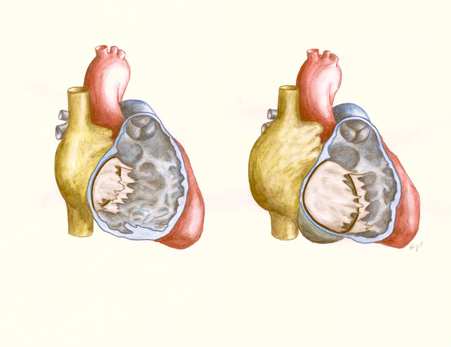
Figure 21. Schematic drawing showing Ebstein’s anomaly of the tricuspid valve. Left: normal heart with openend right ventricle. Right: Ebstein’s anomaly with displacement of the septal and posterior tricuspid leaflet, leading to atrialisation of a significant part of the right ventricle. |
|---|---|
| Source |
illustration by dr. J.P.M. Hamer |
| Date | |
| Author |
illustration by dr. J.P.M. Hamer |
| Permission |
with permission |
File history
Click on a date/time to view the file as it appeared at that time.
| Date/Time | Thumbnail | Dimensions | User | Comment | |
|---|---|---|---|---|---|
| current | 16:52, 1 February 2012 | | 1,462 × 1,126 (1.44 MB) | Nja (talk | contribs) | {{Information |Description=Figure 21. Schematic drawing showing Ebstein’s anomaly of the tricuspid valve. Left: normal heart with openend right ventricle. Right: Ebstein’s anomaly with displacement of the septal and posterior tricuspid leaflet, leadin |
You cannot overwrite this file.
File usage
The following page uses this file: