File:Figure 12.jpg

Original file (4,606 × 4,000 pixels, file size: 1.99 MB, MIME type: image/jpeg)
| Description |
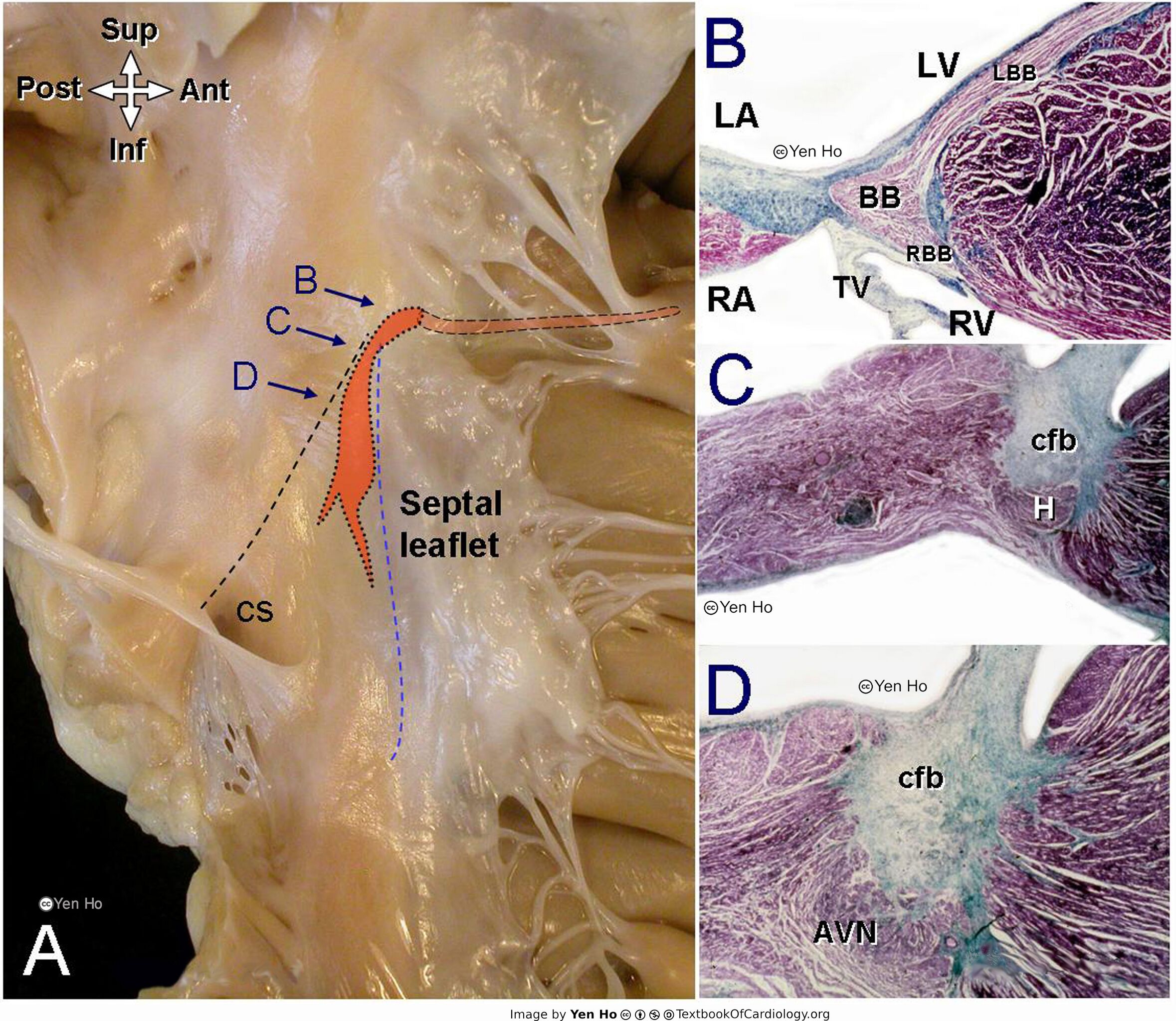
A. This view of the right atrium and right ventricle shows the anterior and posterior borders of the triangle of Koch (broken lines) that mark location of the atrioventricular node and bundle (orange shapes). The arrows B, C, D indicate the cuts made through the conduction system as shown on the histologic sections.
|
|---|---|
| Source |
provided by S. Yen Ho, PhD FRCPath FESC FHEA, Royal Brompton Hospital, UK |
| Date |
2012 |
| Author |
S. Yen Ho, PhD FRCPath FESC FHEA, Royal Brompton Hospital, UK |
| Permission |
File history
Click on a date/time to view the file as it appeared at that time.
| Date/Time | Thumbnail | Dimensions | User | Comment | |
|---|---|---|---|---|---|
| current | 11:49, 18 May 2012 | | 4,606 × 4,000 (1.99 MB) | NiloferT (talk | contribs) |
You cannot overwrite this file.
File usage
The following page uses this file: