File:Figure 11.jpg
Original file (4,961 × 3,247 pixels, file size: 1.43 MB, MIME type: image/jpeg)
| Description |
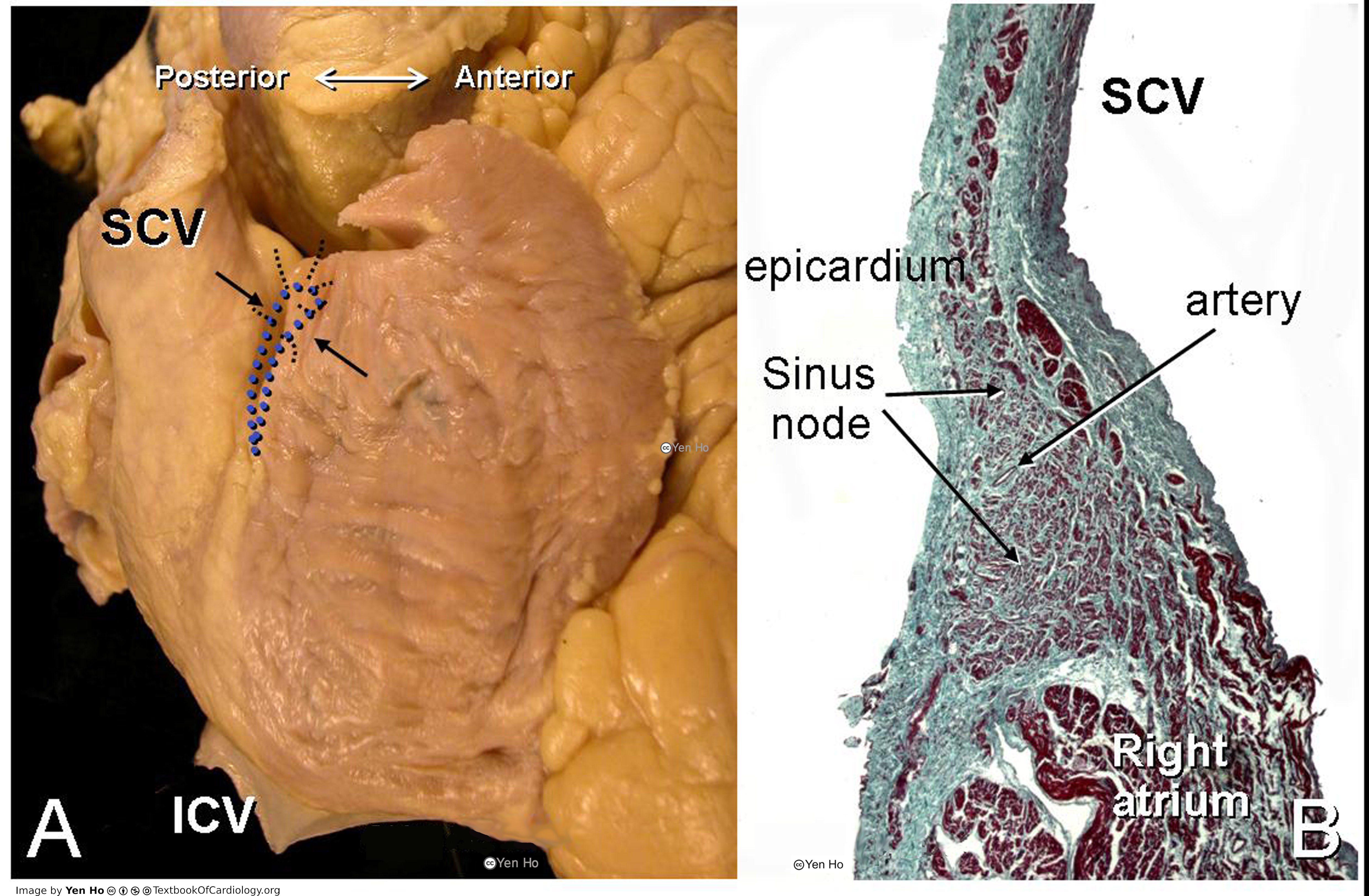
A. The sinus node (dotted shape) is superimposed onto the terminal groove in this picture of the right atrium viewed from the right side. The arrows indicate the sectioning plane of the histological section shown in B.
|
|---|---|
| Source |
provided by S. Yen Ho, PhD FRCPath FESC FHEA, Royal Brompton Hospital, UK |
| Date |
2012 |
| Author |
S. Yen Ho, PhD FRCPath FESC FHEA, Royal Brompton Hospital, UK |
| Permission |
File history
Click on a date/time to view the file as it appeared at that time.
| Date/Time | Thumbnail | Dimensions | User | Comment | |
|---|---|---|---|---|---|
| current | 11:30, 18 May 2012 | | 4,961 × 3,247 (1.43 MB) | NiloferT (talk | contribs) |
You cannot overwrite this file.
File usage
The following page uses this file: