Uploads by NiloferT
Jump to navigation
Jump to search
This special page shows all uploaded files.
| Date | Name | Thumbnail | Size | Description | Versions |
|---|---|---|---|---|---|
| 21:38, 28 November 2013 | Heart4.JPG (file) |  |
56 KB | 1 | |
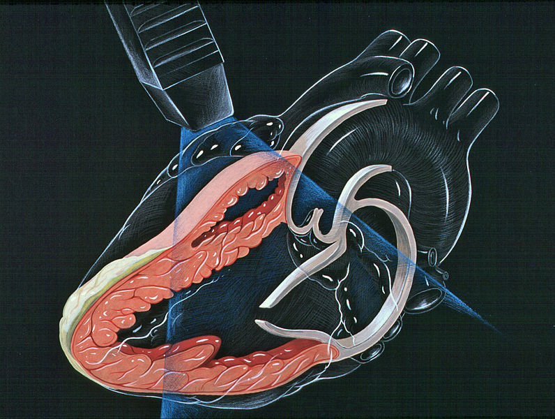
| 18:29, 9 October 2012 | Heart bicuspid aortic valve.svg (file) |  |
28 KB | Description: Heart bicuspid aortic valve anatomy Date: 23 December 2006 Author: Patrick J. Lynch, medical illustrator Creative Commons Attribution 2.5 License 2006 | 1 |
| 18:42, 18 October 2012 | Heart lpla echocardiography diagram.jpg (file) |  |
133 KB | Description: Heart normal LPLA left parasternal long axis echocardiography view Date: 23 December 2006 Author: Patrick J. Lynch, medical illustrator | 1 |
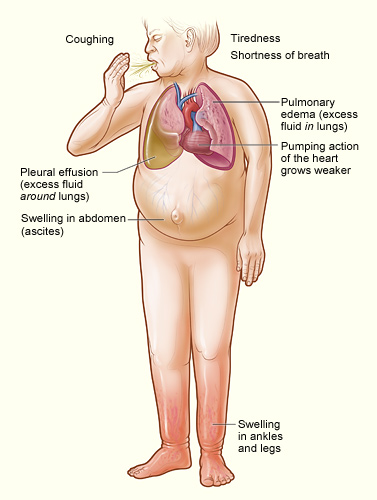
| 13:41, 17 January 2013 | Heartfailure.jpg (file) |  |
55 KB | Description: The illustration shows the major signs and symptoms of heart failure. Date: Originally uploaded to en.wikipedia on 22 September 2008. Source: http://www.nhlbi.nih.gov/health/dci/Diseases/Hf/HF_SignsAndSymptoms.html; transferred from en.w... | 1 |
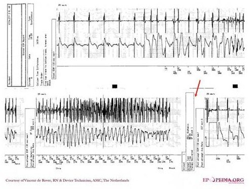
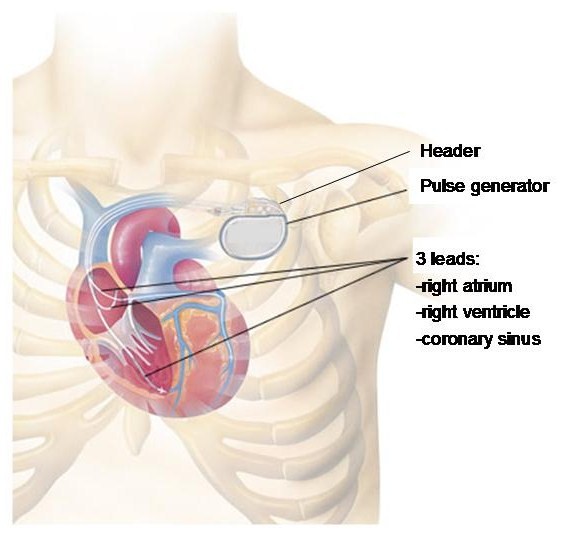
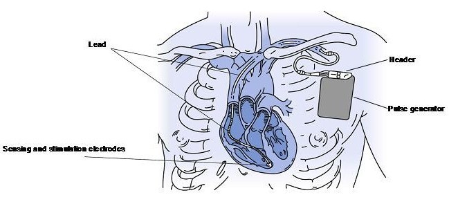
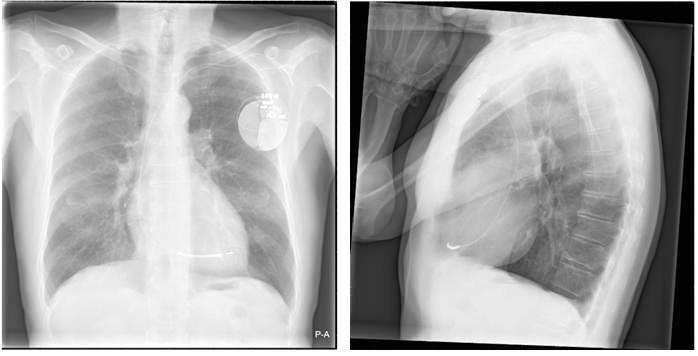
| 02:53, 6 August 2012 | ICD.jpg (file) |  |
67 KB | 1 | |
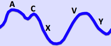
| 14:56, 28 November 2012 | Jugular Venous Pulse.png (file) |  |
16 KB | Description: The Jugular Venous Pressure Waveform Date:13 December 2011 Author Ecgtocardiology | 1 |
| 18:50, 18 October 2012 | LeftVentricleShortAxis.gif (file) |  |
64 KB | Description: Short axis view of left ventricle of heart Date: 10 July 1999 Source:http://www.yale.edu/imaging/echo_atlas/views/short_axis_lv.html Author: Patrick J. Lynch and C. Carl Jaffe | 1 |
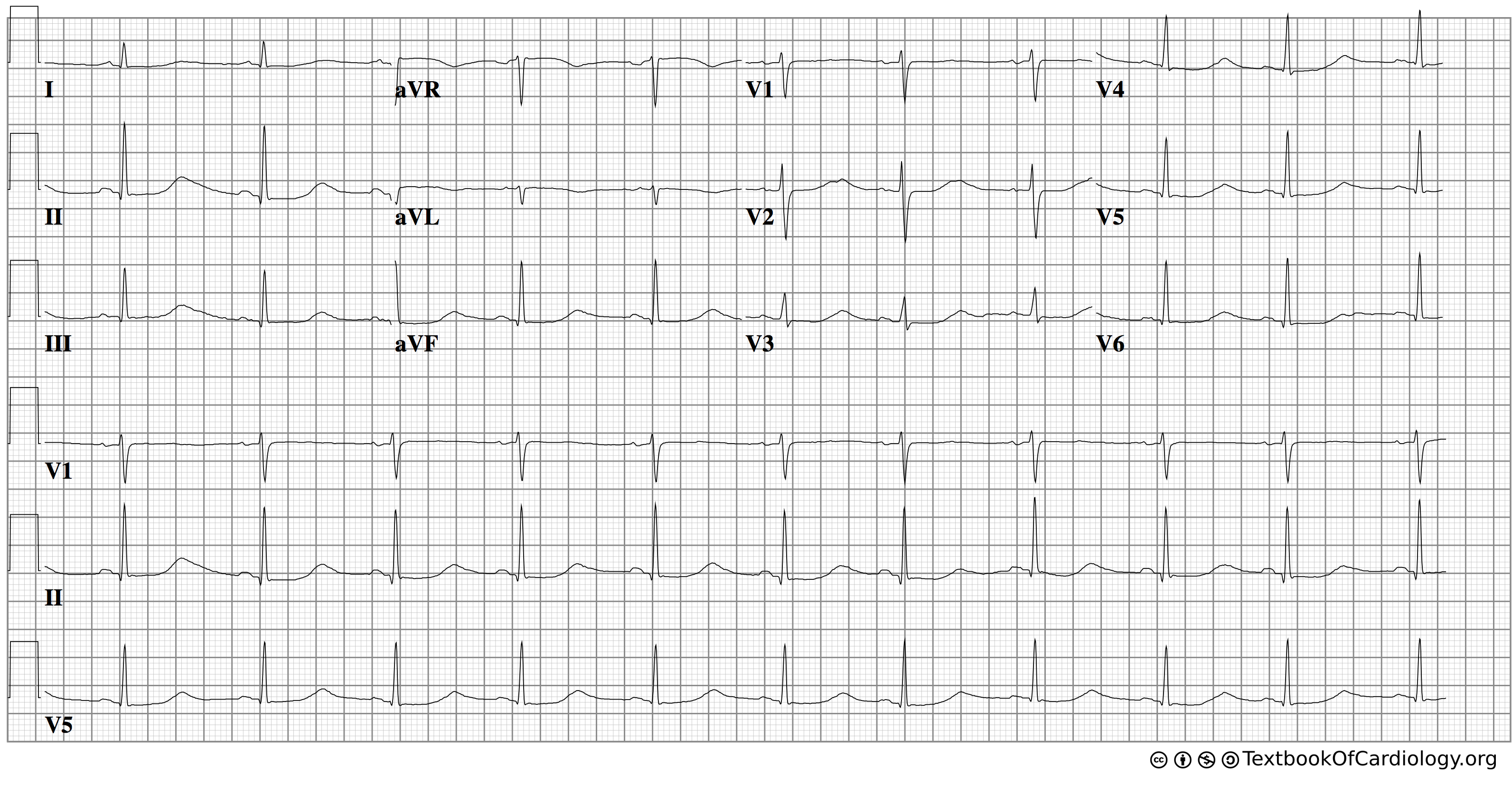
| 13:54, 12 December 2012 | Lqts1.png (file) |  |
417 KB | 2 | |
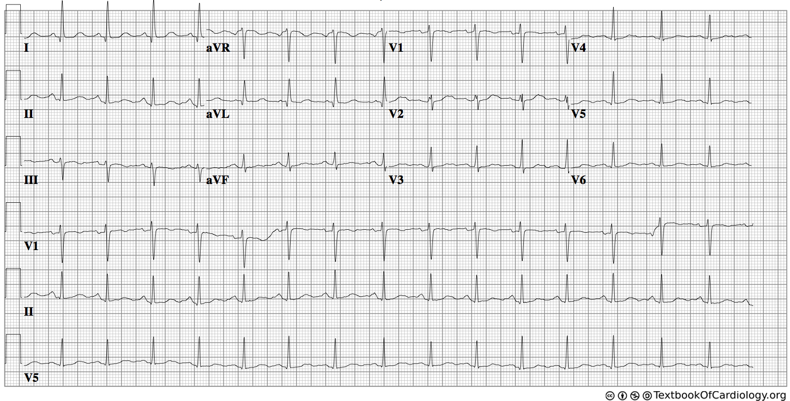
| 14:04, 12 December 2012 | Lqts2.png (file) |  |
309 KB | 3 | |
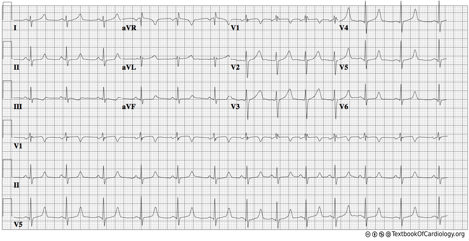
| 20:05, 12 December 2012 | Lqts3.png (file) |  |
316 KB | 4 | |
| 00:08, 4 January 2013 | Main symptoms of diabetes.png (file) |  |
863 KB | 2 | |
| 20:04, 4 October 2012 | Myocardi1.jpg (file) |  |
61 KB | 1 | |
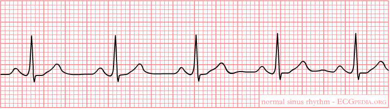
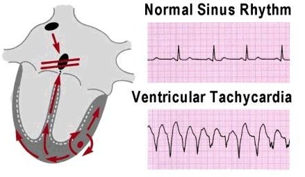
| 23:31, 17 October 2012 | Nsr.png (file) |  |
11 KB | 1 | |
| 04:52, 1 July 2012 | Orthostatic.JPG (file) |  |
25 KB | 1 | |
| 18:41, 29 November 2012 | P1.jpg (file) |  |
13 KB | 1 | |
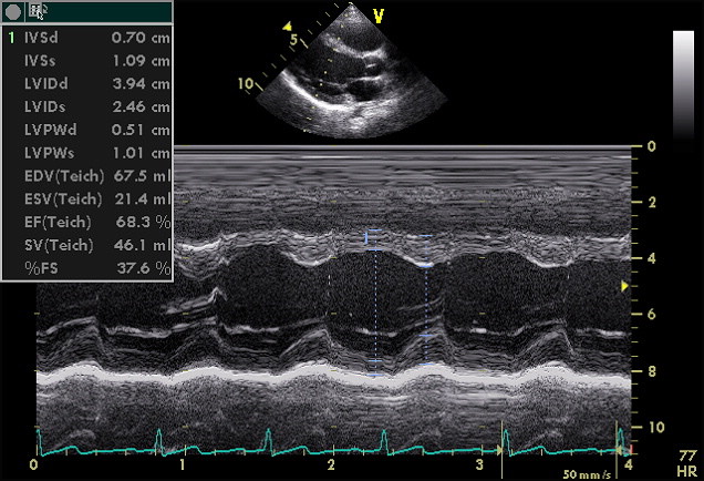
| 18:58, 18 October 2012 | PLAX Mmode.jpg (file) |  |
93 KB | Description: Echocardiogram in the parasternal long-axis view, showing a measurement of the heart's left ventricle Date: May, 2005 Author: Ekko (Uploaded Kjetil Lenes, who made the picture. It is released into the public domain.) | 1 |
| 00:14, 6 August 2012 | Paced2.gif (file) |  |
7 KB | Reverted to version as of 23:31, 5 August 2012 | 3 |
| 04:47, 1 July 2012 | Pathophysiology.JPG (file) |  |
30 KB | 1 | |
| 18:30, 9 October 2012 | Pulmonary valve stenosis.svg (file) |  |
59 KB | Description: The diagram shows a healthy heart and one suffering from Pulmonary valve stenosis. Date: 12 June 2006 Author: Mariana Ruiz LadyofHats | 1 |
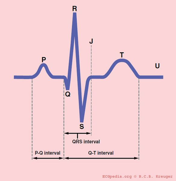
| 18:47, 18 October 2012 | QRSwaves.jpg (file) |  |
20 KB | Description: The different waves and intervals of the ECG (P, PQ, QRS, QT, ST) Date: 2007 Author: Rob Kreuger, medical illustrator, AMC, The Netherlands | 1 |
| 03:35, 6 August 2012 | RedTh.jpg (file) | 31 KB | 1 | ||
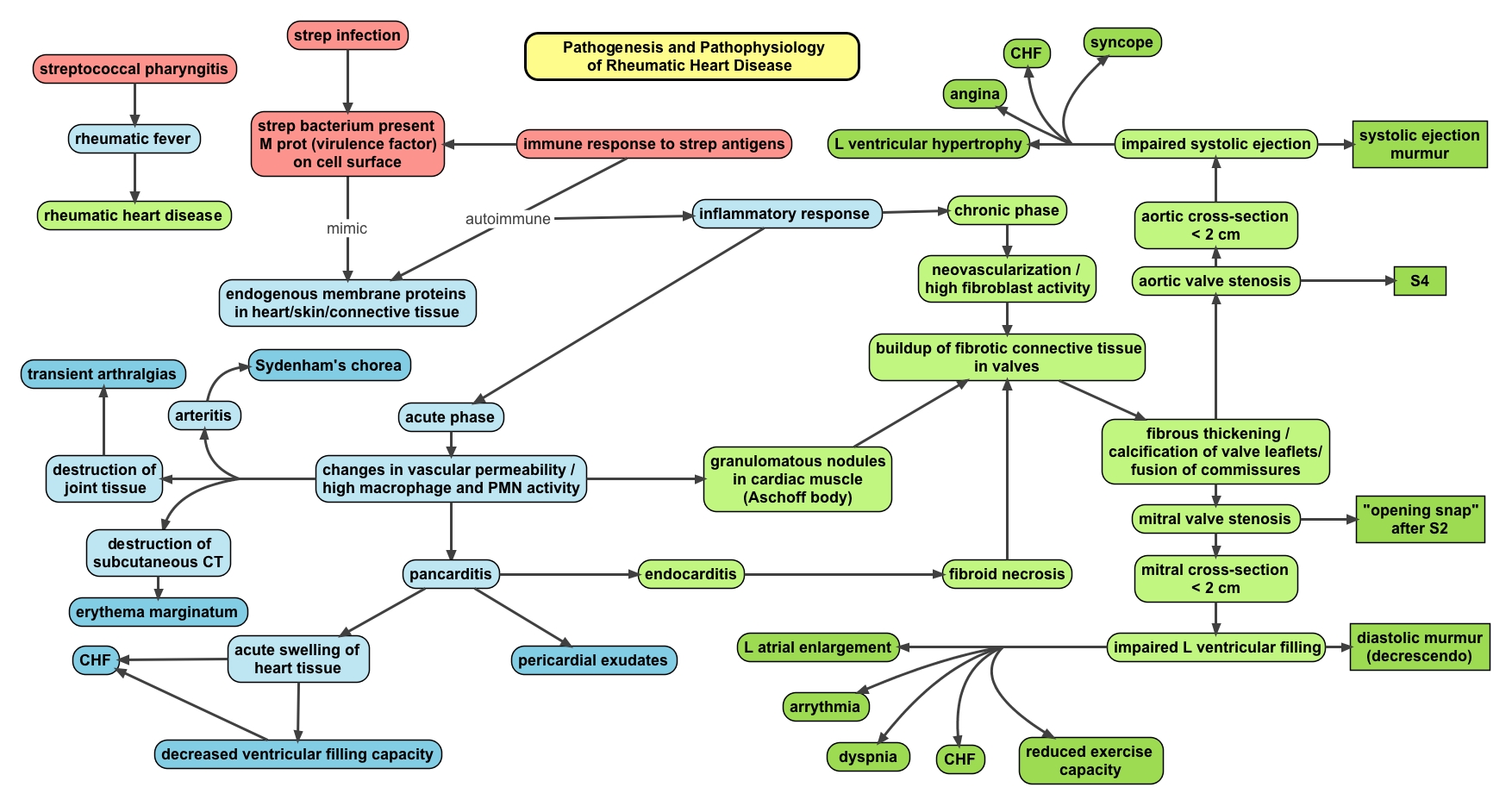
| 15:51, 17 May 2013 | Rheum.heart.disease.jpeg (file) |  |
586 KB | Description: Pathophysiology map of rheumatic fever and rheumatic heart disease. Author: Oxynthes | 1 |
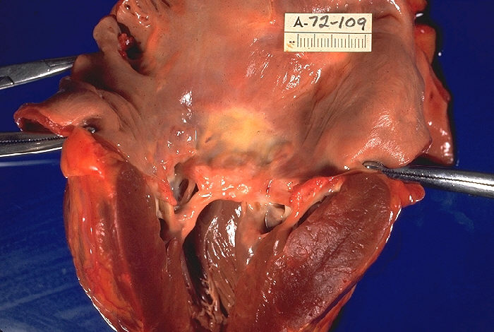
| 15:53, 17 May 2013 | Rheumatic heart disease, gross pathology 20G0013 lores.jpg (file) |  |
80 KB | Description: "Gross pathology of rheumatic heart disease. Left ventricle has been cut open to display characteristic severe thickening of mitral valve, thickened chordae tendineae, and hypertrophied left ventricular myocardium. Autopsy." Date: 1972 Sou... | 1 |
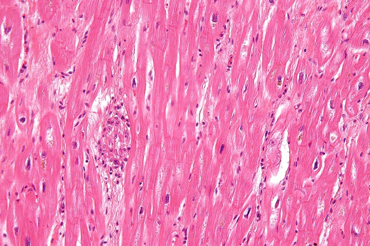
| 18:32, 17 May 2013 | Rheumatic heart disease - 3a - very high mag.jpg (file) |  |
351 KB | Description: Very high magnification micrograph of rheumatic heart disease. H&E stain. It is due to Streptococcus pyogenes. Microscopic findings include Anitschkow cells (also known as caterpillar cells), and Aschoff bodies. Anitschkow cells are though... | 1 |
| 20:38, 17 May 2013 | Rheumatic heart disease world map - DALY - WHO2004.svg (file) |  |
1.45 MB | Description: Age-standardised disability-adjusted life year (DALY) rates from Rheumatic heart disease by country (per 100,000 inhabitants). Source: Vector map from BlankMap-World6, compact.svg by Canuckguy et al. Data from Death and DALY estimates for ... | 1 |
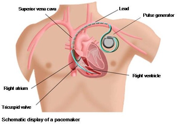
| 22:23, 5 August 2012 | Schematic.jpg (file) |  |
54 KB | 1 | |
| 17:54, 5 August 2012 | SchematicImg.jpg (file) |  |
50 KB | 2 | |
| 22:47, 5 August 2012 | Schematicpic.jpg (file) |  |
53 KB | 2 | |
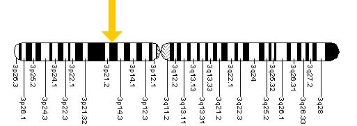
| 09:25, 9 October 2012 | Scn5a.jpg (file) |  |
17 KB | 1 | |
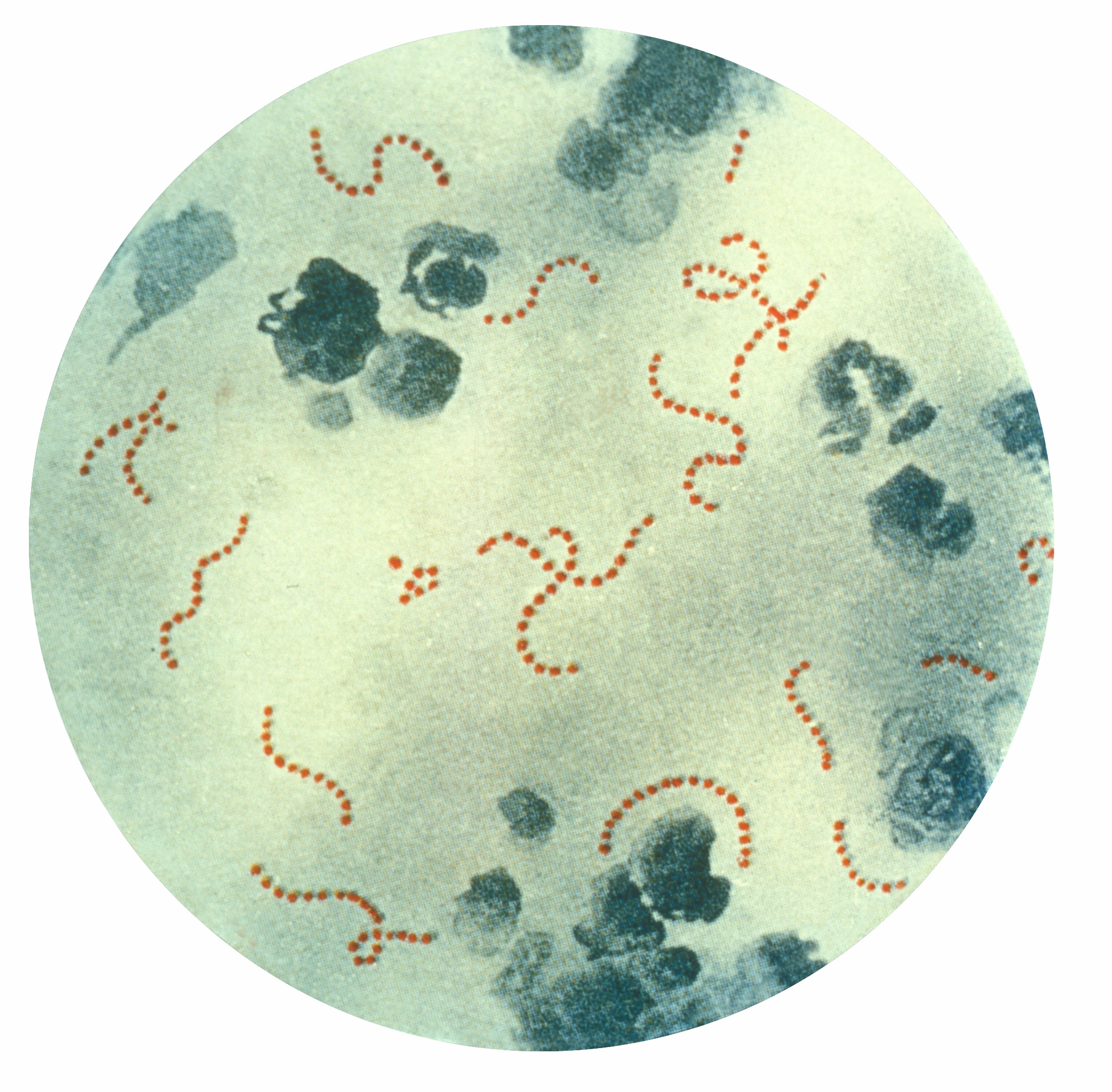
| 16:08, 17 May 2013 | Streptococcus pyogenes 01.jpg (file) |  |
1.01 MB | Description: Photomicrograph of Streptococcus pyogenes bacteria, 900x Mag. A pus specimen, viewed using Pappenheim's stain. Last century, infections by S. pyogenes claimed many lives especially since the organism was the most important cause of puerper... | 2 |
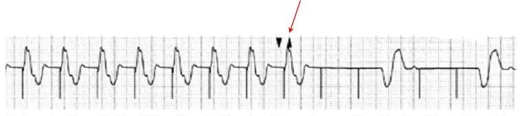
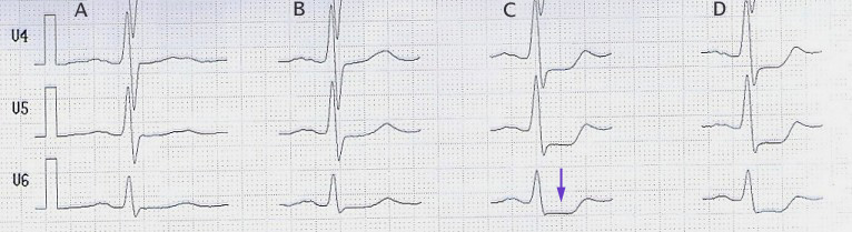
| 23:23, 17 May 2013 | StressECG STDepression.jpg (file) | 72 KB | Description: Belastungs-EKG mit ST-Senkung (Pfeil) ab 100 W (Spalte C) stress-ecg with st-segment-depression (arrow) beginning at 100 W (column C) Date: published 10. Jan. 2006 Source: selbst abgeleitet/own recording Author: J. Heuser JHeuser | 1 | |
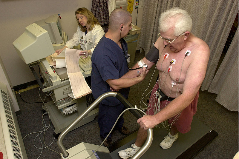
| 18:25, 18 October 2012 | Stress test.jpg (file) |  |
108 KB | Description: Stock footage taken at Beaumont Hospital. 14:18, 28 October 2006(UTC) Date: 2006-10-28 Author: Blue0ctane at en.wikipedia | 1 |
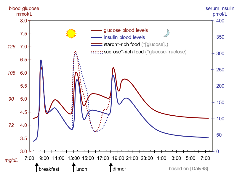
| 21:36, 23 December 2012 | Suckale08 fig3 glucose insulin day.png (file) |  |
80 KB | Description: Idealized curves of human blood glucose and insulin concentrations during the course of a day containing three meals; in addition, effect of sugar-rich meal is highlighted. Source: Solimena Lab and Review Suckale Solimena 2008 Frontiers in... | 1 |
| 03:18, 6 January 2013 | Swe.jpg (file) |  |
57 KB | 3 | |
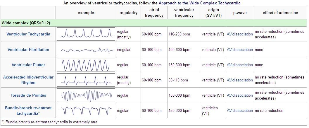
| 20:29, 6 July 2012 | TableVent.jpg (file) |  |
126 KB | 1 | |
| 19:28, 6 July 2012 | Vent1.jpg (file) |  |
39 KB | 1 | |
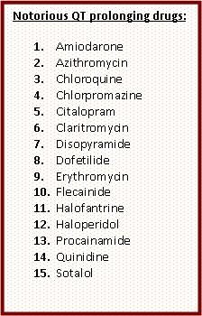
| 20:32, 6 July 2012 | VentDrg.jpg (file) |  |
45 KB | 1 | |
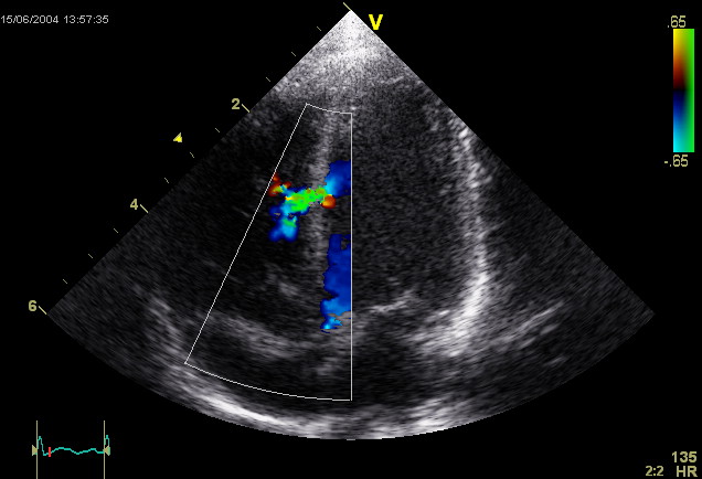
| 23:48, 17 October 2012 | Ventricular Septal Defect.jpg (file) |  |
54 KB | This is an ultrasound picture of the heart, an echocardiogram. It depicts a ventricular septal defect. Author: Kjetil Lenes. | 1 |
| 20:14, 5 August 2012 | Xthorax.jpg (file) |  |
44 KB | 1 |