Uploads by NiloferT
Jump to navigation
Jump to search
This special page shows all uploaded files.
| Date | Name | Thumbnail | Size | Description | Versions |
|---|---|---|---|---|---|
| 12:43, 20 May 2012 | Figure 5.jpg (file) |  |
815 KB | 1 | |
| 10:46, 18 May 2012 | Figure 4.jpg (file) |  |
566 KB | 1 | |
| 10:40, 18 May 2012 | Figure 3.jpg (file) |  |
691 KB | 1 | |
| 10:36, 18 May 2012 | Figure 2.jpg (file) |  |
1.25 MB | 1 | |
| 12:48, 20 May 2012 | Figure 13.jpg (file) |  |
1.48 MB | 1 | |
| 11:49, 18 May 2012 | Figure 12.jpg (file) |  |
1.99 MB | 1 | |
| 11:30, 18 May 2012 | Figure 11.jpg (file) |  |
1.43 MB | 1 | |
| 11:25, 18 May 2012 | Figure 10.jpg (file) |  |
1.3 MB | 1 | |
| 10:21, 18 May 2012 | Figure1.jpg (file) |  |
1.06 MB | 1 | |
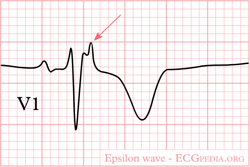
| 21:45, 31 October 2012 | Epsilon wave.png (file) |  |
14 KB | 1 | |
| 03:46, 6 August 2012 | ECGT.jpg (file) |  |
57 KB | 1 | |
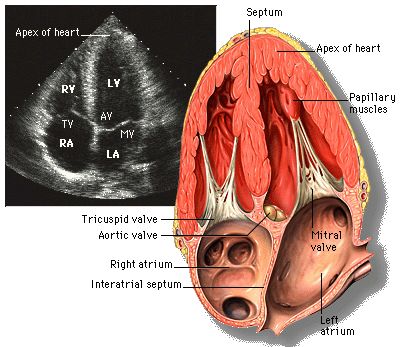
| 18:23, 9 October 2012 | Diagram of the human heart (valves improved).svg (file) | .svg) |
27 KB | Source:http://commons.wikimedia.org/wiki/Image:Diagram_of_the_human_heart_%28cropped%29.svg | 2 |
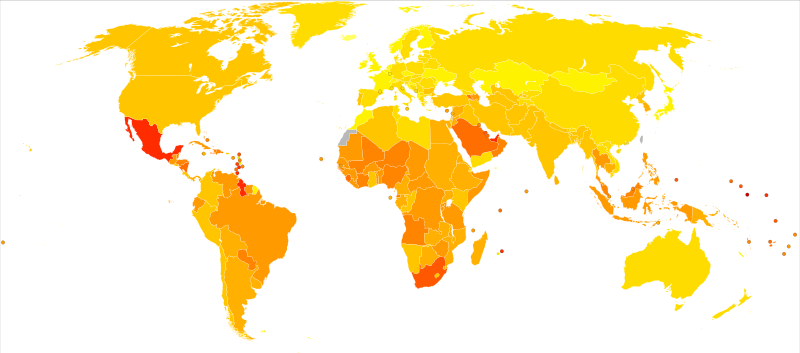
| 00:24, 4 January 2013 | Diabetes world map - 2000.png (file) |  |
1.45 MB | 2 | |
| 23:11, 23 December 2012 | Diabetes mellitus world map - DALY - WHO2004.svg (file) |  |
78 KB | Description: Age-standardised disability-adjusted life year (DALY) rates from Diabetes mellitus by country (per 100,000 inhabitants). Date: 11 January 2010 Source: Vector map from BlankMap-World6, compact.svg by Canuckguy et al. Data from Death and DAL... | 1 |
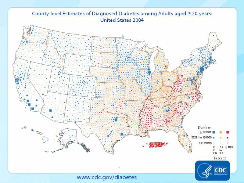
| 22:52, 23 December 2012 | Diabetes County level estimates 2004-2009.gif (file) |  |
363 KB | Description: An animated map of the United States showing the prevalence of diabetes from 2004-2009. Date: 12 August 2012 Source: http://www.cdc.gov/obesity/data/adult.html from http://apps.nccd.cdc.gov/DDT_STRS2/NationalDiabetesPrevalenceEstimates.asp... | 1 |
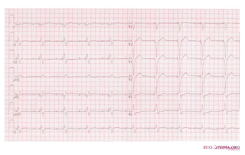
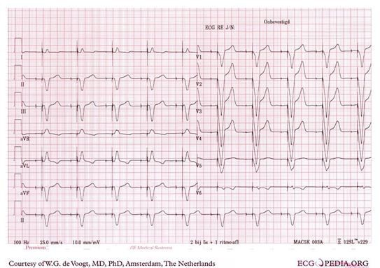
| 00:20, 6 August 2012 | Ddd paced 12lead.jpg (file) |  |
73 KB | 1 | |
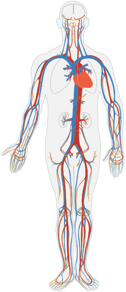
| 18:42, 29 November 2012 | Circulatory System no tags.svg (file) |  |
104 KB | 2 | |
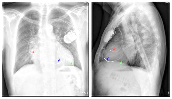
| 19:52, 5 August 2012 | ChestXray.jpg (file) |  |
46 KB | 2 | |
| 09:29, 9 October 2012 | Brugada syndrome type2 example2.jpg (file) |  |
677 KB | 1 | |
| 09:26, 9 October 2012 | Brugada syndrome type2 example1.png (file) |  |
363 KB | 1 | |
| 09:31, 9 October 2012 | Brugada syndrome type1 example6.jpg (file) |  |
970 KB | 1 | |
| 09:25, 9 October 2012 | Brugada syndrome type1 example5.png (file) |  |
364 KB | 1 | |
| 09:24, 9 October 2012 | Brugada syndrome type1 example4.png (file) |  |
364 KB | 1 | |
| 09:22, 9 October 2012 | Brugada syndrome type1 example3.png (file) |  |
63 KB | 1 | |
| 09:26, 9 October 2012 | Brugada syndrome type1 example2.png (file) |  |
348 KB | 1 | |
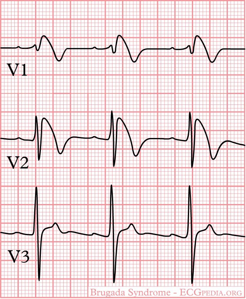
| 09:25, 9 October 2012 | Brugada syndrome type1 example1.png (file) |  |
370 KB | 1 | |
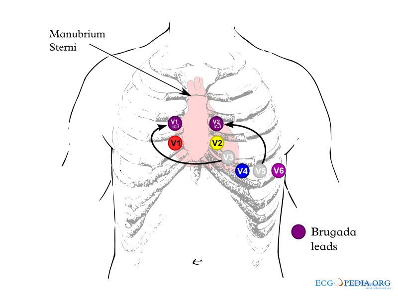
| 09:21, 9 October 2012 | Brugada lead placement.png (file) |  |
84 KB | 1 | |
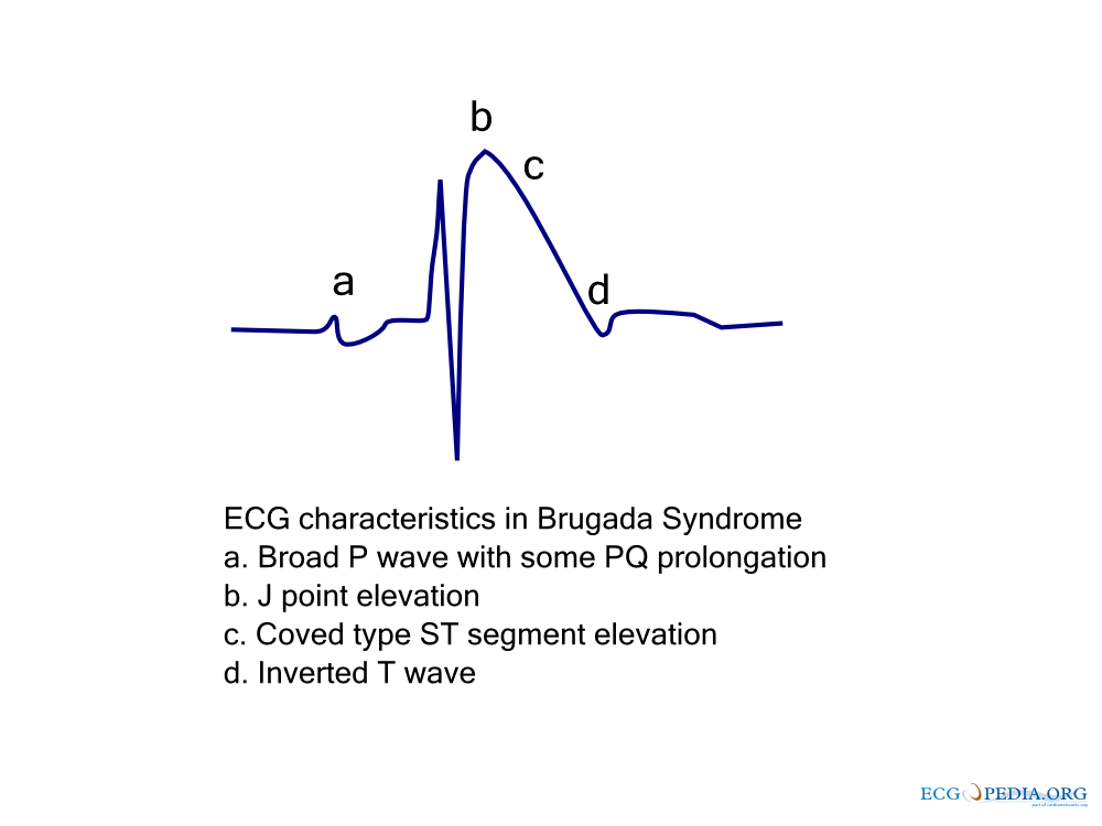
| 09:21, 9 October 2012 | Brugada ecg characteristics.png (file) |  |
39 KB | 1 | |
| 09:21, 9 October 2012 | Brugada.png (file) |  |
29 KB | 1 | |
| 09:21, 9 October 2012 | Brugada.jpg (file) |  |
13 KB | 1 | |
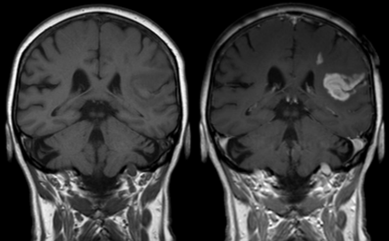
| 14:21, 17 January 2013 | Bluthirnschranke nach Infarkt nativ und KM.png (file) |  |
549 KB | Description: Defect of the blood-brain barrier after stroke shown in MRI. T1-weighted images, left image without right image with contrast medium administration. Deutsch: Störung der Blut-Hirn-Schranke nach einem ischämischen Hirninfarkt im Stromgeb... | 1 |
| 23:41, 23 December 2012 | Blue circle for diabetes.svg.png (file) |  |
8 KB | Description: The blue circle is the global symbol for diabetes, introduced by the International Diabetes Federation with the aim of giving diabetes a common identity, supporting existing efforts to raise awareness of diabetes and placing the diabetes e... | 1 |
| 19:19, 6 July 2012 | Bb reentry small.svg (file) | 52 KB | 5 | ||
| 04:41, 6 August 2012 | AtrialCap.jpg (file) |  |
75 KB | 1 | |
| 21:50, 31 October 2012 | Arvdhart.png (file) |  |
964 KB | 1 | |
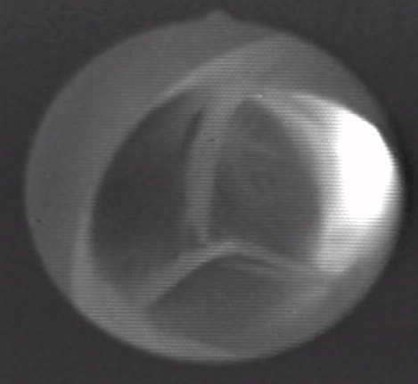
| 18:38, 18 October 2012 | Apical 4 chamber view.gif (file) |  |
72 KB | Description: Apical four chamber view of heart Date: 10 July 1999 Source: http://www.yale.edu/imaging/echo_atlas/views/four_chamber.html Author: Patrick J. Lynch and C. Carl Jaffe | 1 |
| 20:23, 9 October 2012 | Aortic valve (1).gif (file) | .gif) |
2.23 MB | This is a video clip from a living, beating pig heart that was prepared in the laboratory as a working Langendorf preparation. The heart was arrested, connected to the perfusion system and restarted. The working fluid was oxygenated balanced saline sol... | 2 |
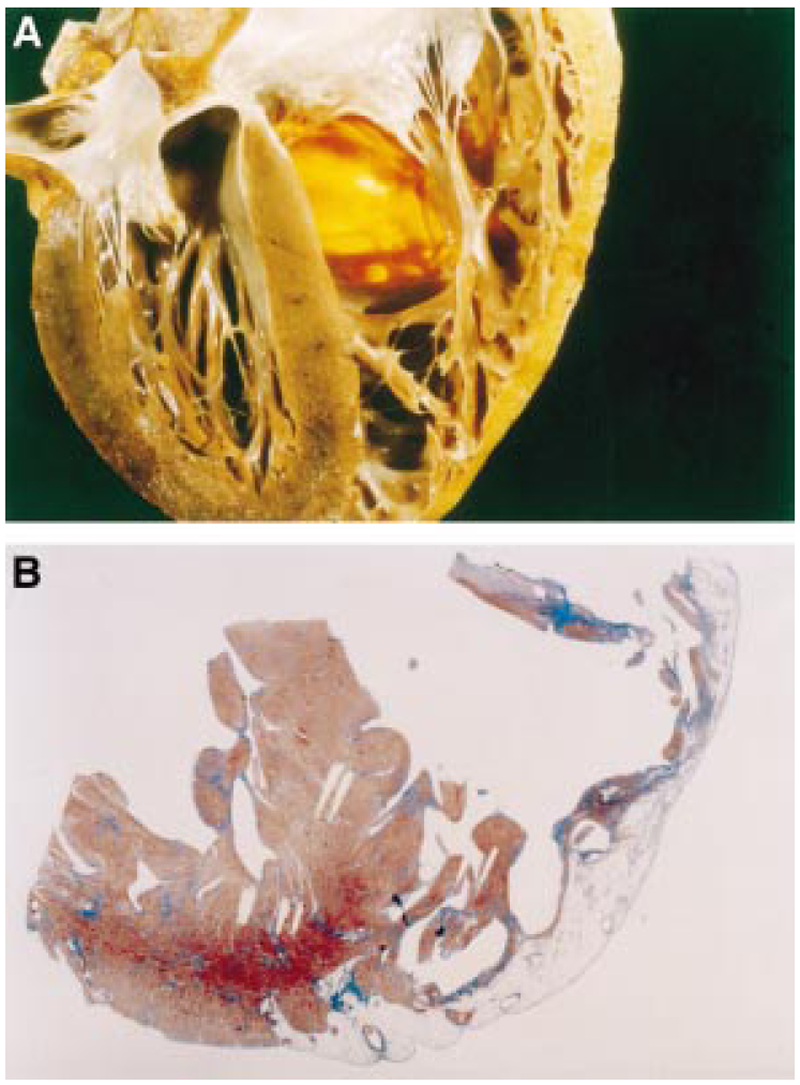
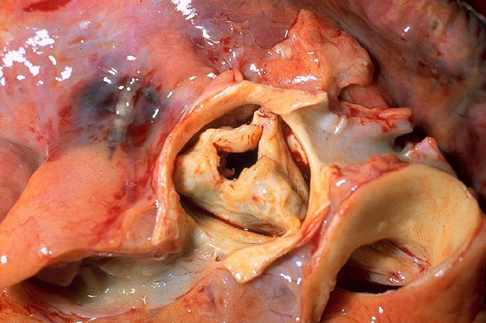
| 18:28, 9 October 2012 | Aortic stenosis rheumatic, gross pathology 20G0014 lores.jpg (file) |  |
92 KB | Description: Gross pathology of rheumatic heart disease: aortic stenosis. Aorta has been removed to show thickened, fused aortic valve leaflets and opened coronary arteries from above. Autopsy. Content Providers(s). Author: CDC/Dr. Edwin P. Ewing, Jr. ... | 1 |
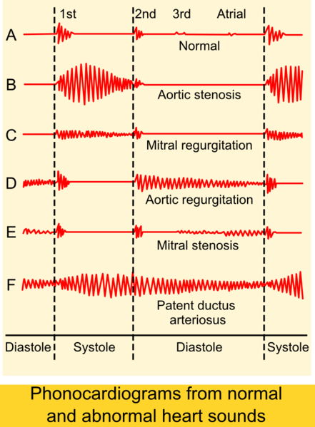
| 18:27, 28 November 2012 | 441px-Phonocardiograms from normal and abnormal heart sounds.png (file) | 125 KB | Description: Phonocardiograms from normal and abnormal heart sounds Author: Madhero88 Source:http://en.wikipedia.org/wiki/File:Phonocardiograms_from_normal_and_abnormal_heart_sounds.png | 1 |