Uploads by Nja
Jump to navigation
Jump to search
This special page shows all uploaded files.
| Date | Name | Thumbnail | Size | Description | Versions |
|---|---|---|---|---|---|
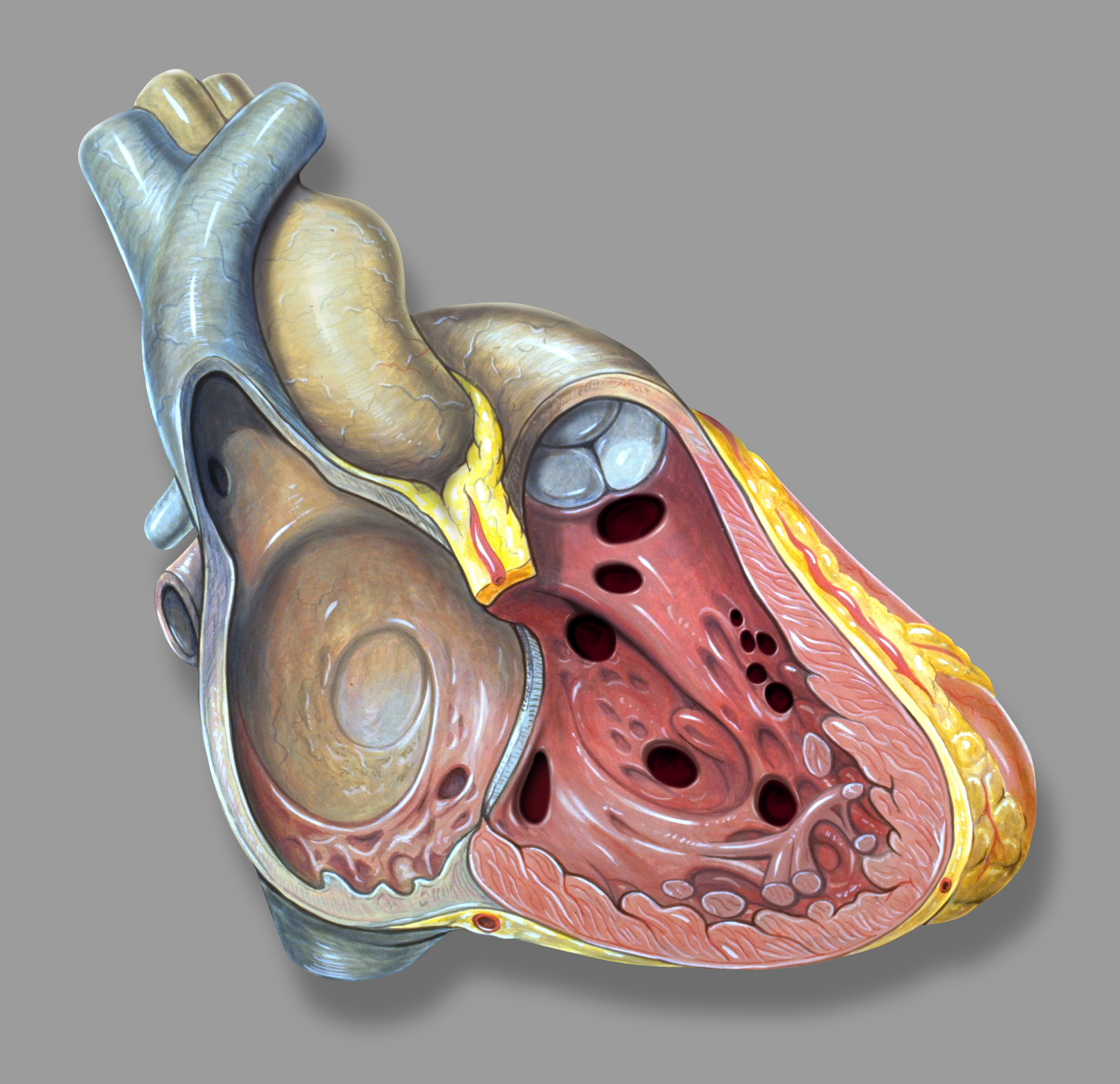
| 14:10, 25 January 2012 | 4. VSD.jpg (file) |  |
1.23 MB | {{Information |Description=Heart from the right side view, showing different locations of ventricular septal defects. |Source=from commons.wikipedia.org |Date=Published: |Author= |Permission= |other_versions= }} | 1 |
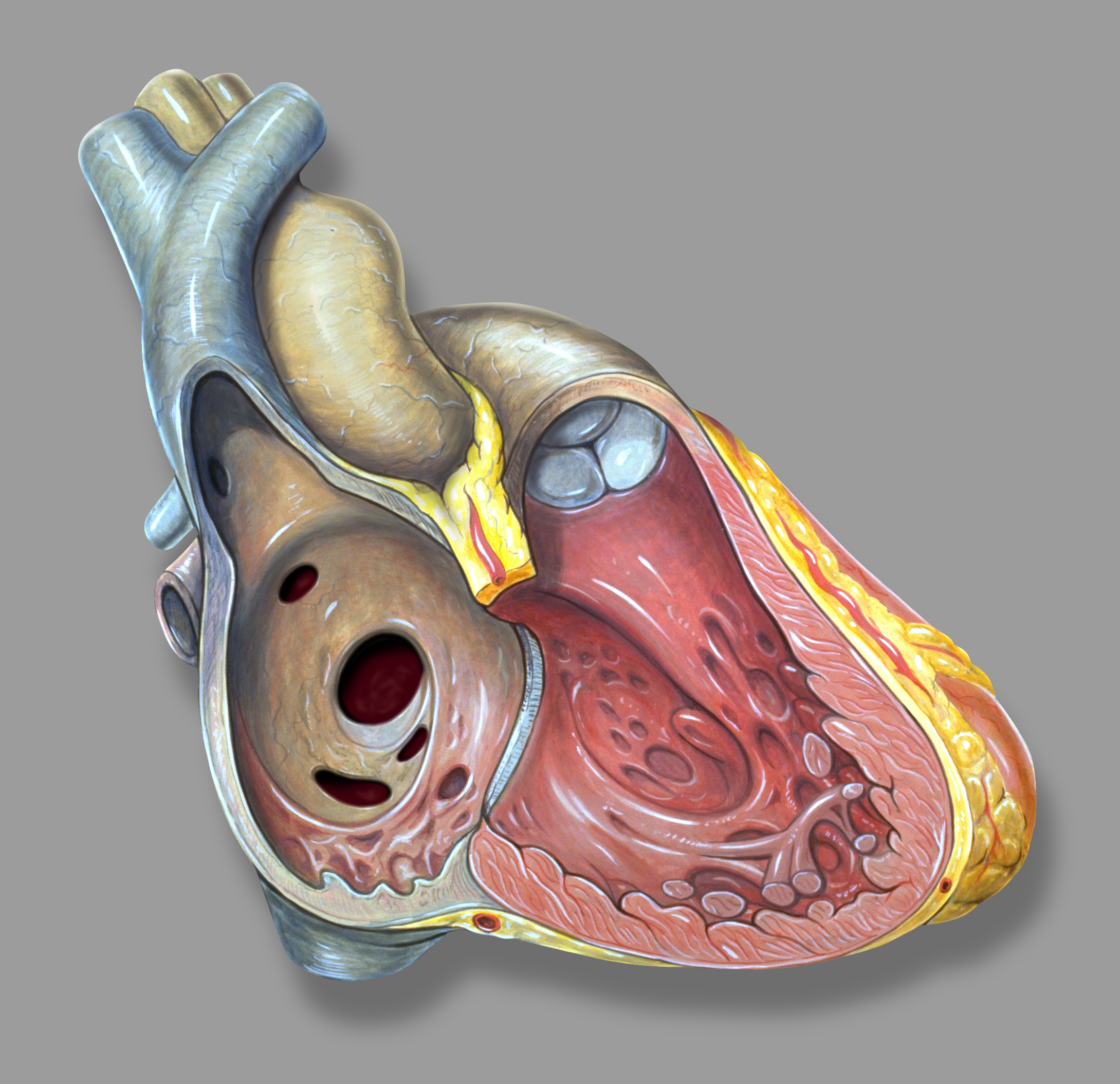
| 23:49, 22 January 2012 | 1. ASD.jpg (file) |  |
1.24 MB | {{Information |Description=Figure 1. Heart from the right side view, showing different locations of atrial septal defects. |Source=from commons.wikipedia.org |Date=Published: |Author= |Permission= |other_versions= }} | 1 |
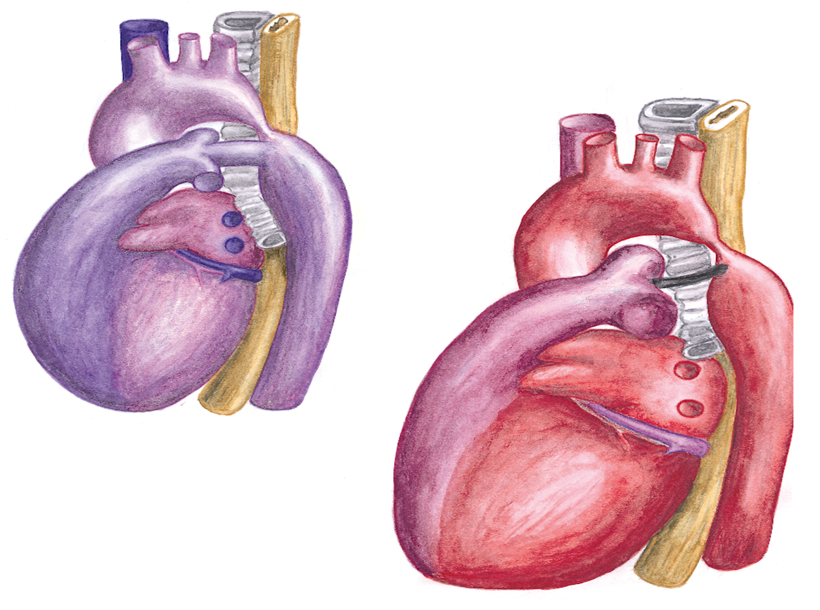
| 16:10, 1 February 2012 | Figure 9. Schematic drawing of the anatomy prenatal and postnatal.png (file) |  |
1.24 MB | {{Information |Description=Figure 9. Schematic drawing of the anatomy prenatal (left) and postnatal (right) in coarctation of the aorta. In the normal situation (without coarctation) only 10 percent of the fetal cardiac output flows through the descending | 1 |
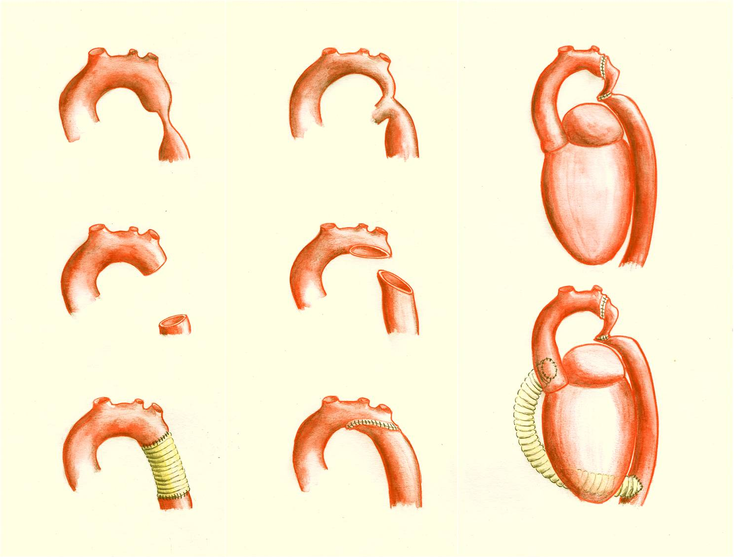
| 16:25, 1 February 2012 | Figure 11. Schematic drawing showing surgical procedures for repair of a coarctation of the aorta.png (file) |  |
1.3 MB | {{Information |Description=Figure 11. Schematic drawing showing surgical procedures for repair of a coarctation of the aorta. Left: an interposition graft. Middle: the extended aortic arch repair. Right: the extra-anatomical bypass. |Source=cillustration | 1 |
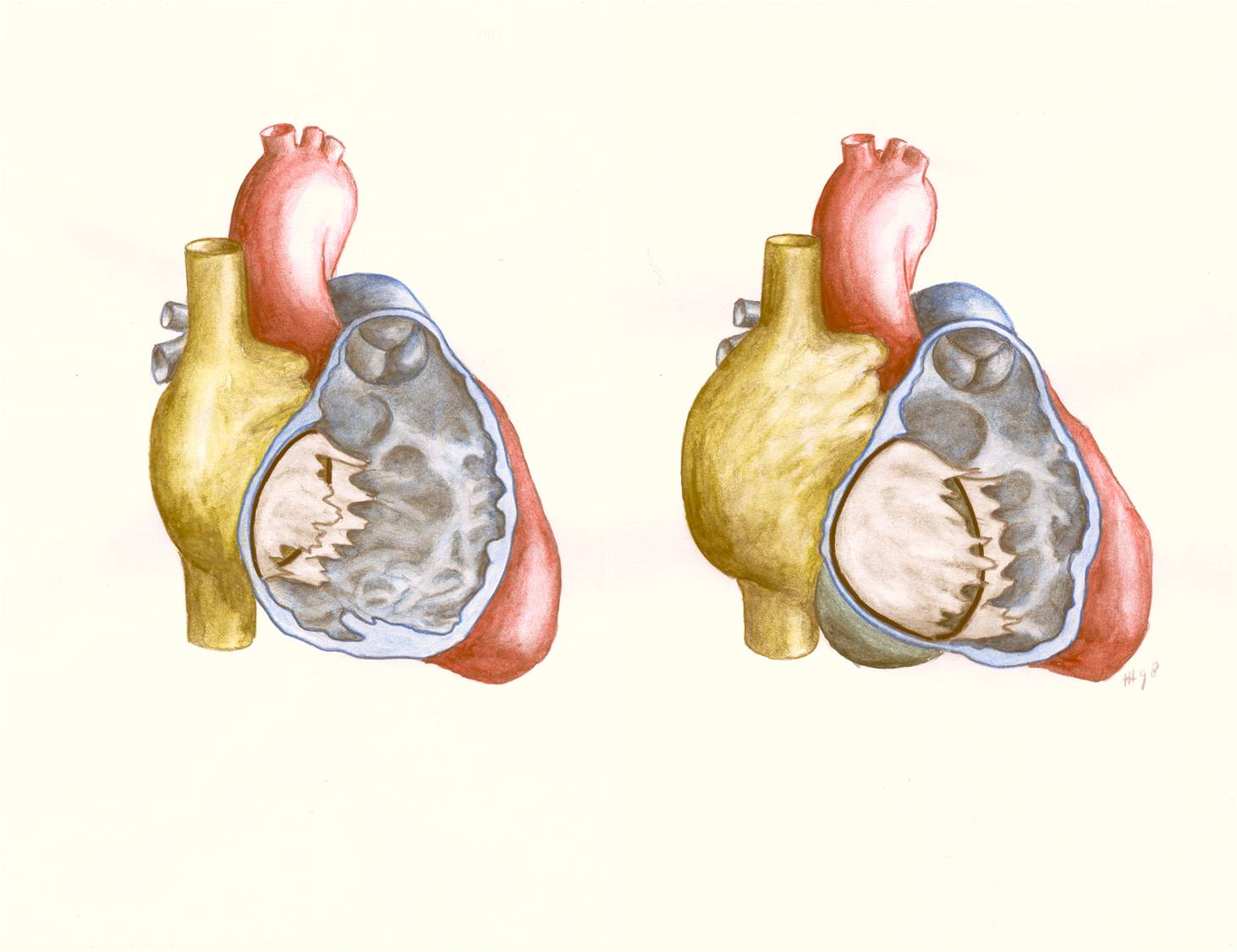
| 16:52, 1 February 2012 | Figure 21. Schematic drawing showing Ebstein’s anomaly of the tricuspid valve.png (file) |  |
1.44 MB | {{Information |Description=Figure 21. Schematic drawing showing Ebstein’s anomaly of the tricuspid valve. Left: normal heart with openend right ventricle. Right: Ebstein’s anomaly with displacement of the septal and posterior tricuspid leaflet, leadin | 1 |
| 14:45, 25 January 2012 | 5. AVSD.PNG (file) |  |
6.65 MB | {{Information |Description=Figure 5. Left: schematic drawing of a normal heart with a normal mitral (M) and tricuspid (T) valve. Middle: a complete AVSD where the mitral and tricuspid valve is replaced by a common valve with a right anterosuperior leaflet | 1 |