Uploads by Drj
Jump to navigation
Jump to search
This special page shows all uploaded files.
| Date | Name | Thumbnail | Size | Description | Versions |
|---|---|---|---|---|---|
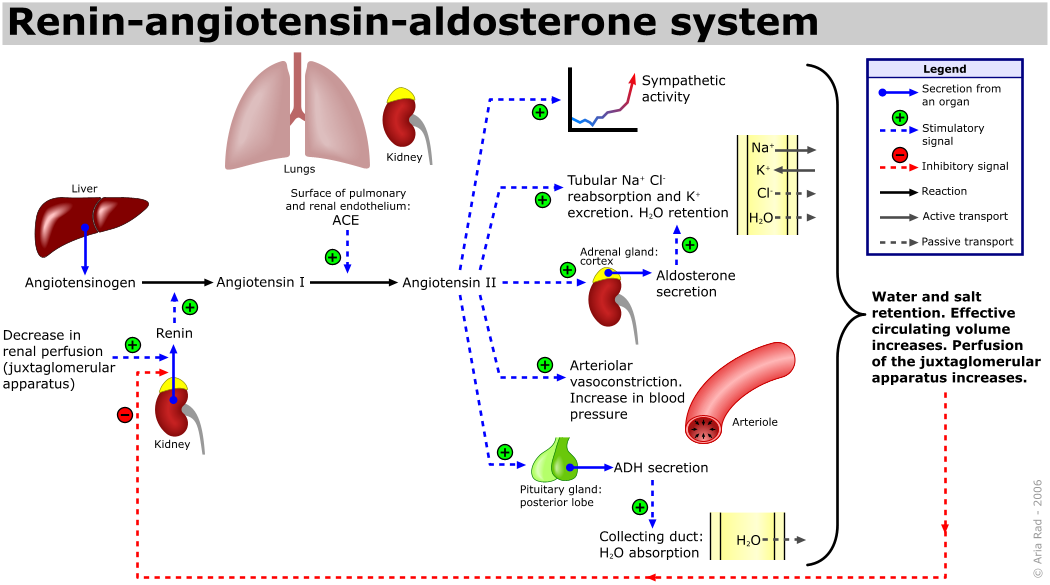
| 08:40, 28 May 2015 | Renin-angiotensin-aldosterone system.png (file) |  |
124 KB | {{Information |description= {{en|1=The renin-angiotensin system (RAS) or the renin-angiotensin-aldosterone system (RAAS) is a great system when it comes to sucking up the great H20. This has proven to change lives of many people and is constantly used... | 2 |
| 20:36, 26 May 2015 | Transesophageal echocardiography diagram.svg (file) | 57 KB | 2 | ||
| 20:29, 26 May 2015 | Heart short axis myocardial segments.svg (file) |  |
17 KB | 2 | |
| 22:13, 29 December 2013 | CPVT.svg (file) |  |
467 KB | 1 | |
| 22:10, 29 December 2013 | Platelet receptors.svg (file) |  |
70 KB | 1 | |
| 21:55, 29 December 2013 | Sympathic parasympathic.svg (file) |  |
1.01 MB | 1 | |
| 19:03, 22 June 2013 | HONConduct792789 s.gif (file) |  |
2 KB | 1 | |
| 18:02, 21 March 2013 | PlaatjesBrS pyramid.svg (file) |  |
28 KB | 2 | |
| 21:23, 21 February 2013 | MEWS.svg (file) |  |
111 KB | 4 | |
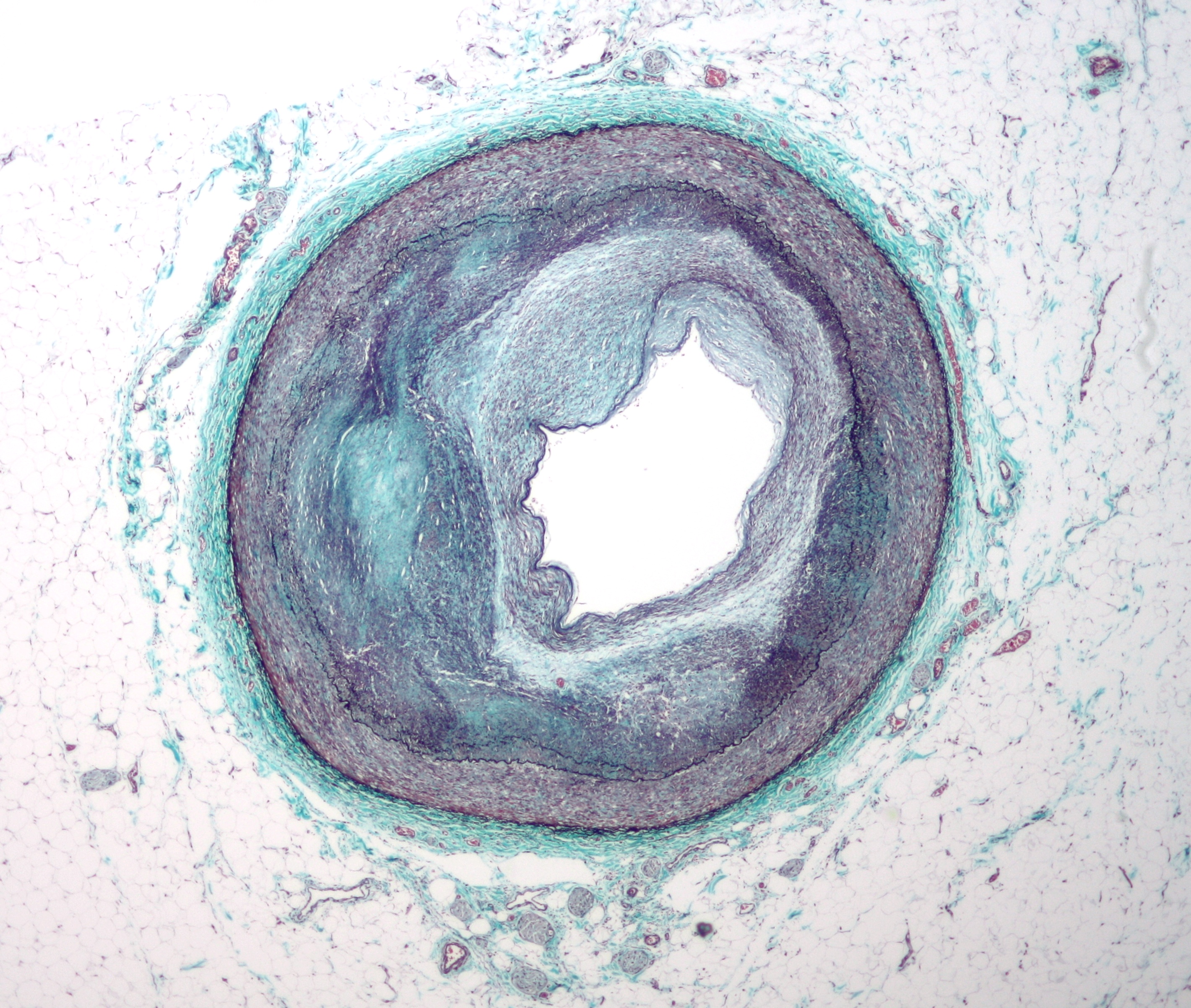
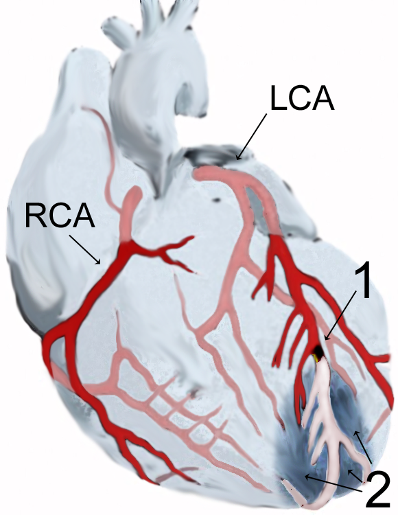
| 14:25, 7 January 2013 | RCA atherosclerosis.jpg (file) |  |
2.05 MB | == {{int:filedesc}} == {{Information |Description={{en|1=Low magnification micrograph of the distal right coronary artery with complex '''atherosclerosis''' and luminal nar... | 1 |
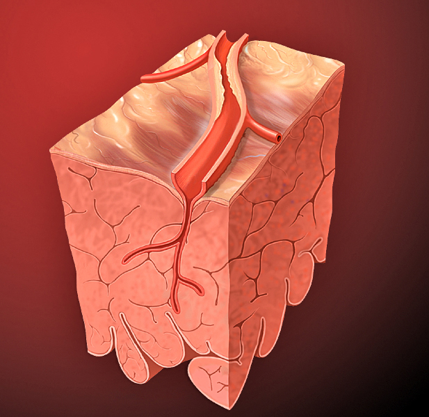
| 20:23, 6 January 2013 | Heart coronary artery.jpg (file) |  |
295 KB | == {{int:filedesc}} == {{Information |Description = Lesion and partial blockage of cornary artery |Source = Patrick J. Lynch, medical illustrator |Date = 2006-12-23 |Author = Patrick J. Lynch, medical illustrator |Permission = Creative Commons Attribut... | 1 |
| 19:59, 3 January 2013 | ALS.svg (file) |  |
28 KB | 3 | |
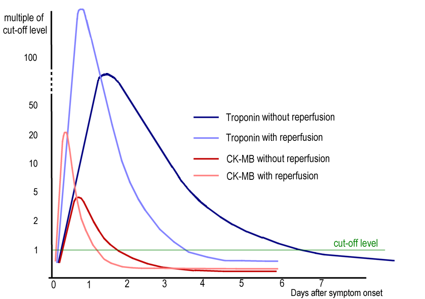
| 20:13, 19 December 2012 | Cardiac markers.png (file) |  |
56 KB | {{Information| |Description = typical changes in CK-MB and cardiac troponin in Acute Myocardial Infarction |Source = self made modified from ACC/AHA Practice Guidelines 2005 p E32 - selbst gemacht abgewandelt von ACC/AHA Practice Guidelines S. E32 |... | 1 |
| 17:06, 18 December 2012 | SR VT.svg (file) |  |
76 KB | 1 | |
| 17:03, 18 December 2012 | OCT ACT.svg (file) |  |
82 KB | 1 | |
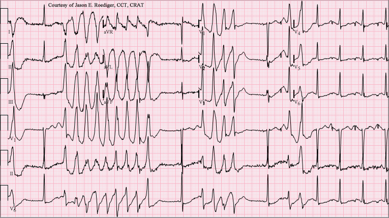
| 20:45, 10 December 2012 | Torsades de Pointes.png (file) |  |
135 KB | 1 | |
| 07:05, 7 December 2012 | Chest pain areas.svg (file) |  |
20 KB | 3 | |
| 12:55, 5 December 2012 | AMI scheme.png (file) |  |
421 KB | {{Information| |Description = myocardial infarction - Myokardinfarkt - scheme |Source = {{own}} |Date = created 19. June 2006 |Author = J. Heuser JHeuser |Permission = {{GFDL-self|migration=relicense}} |other_versions = }} | 1 |
| 09:15, 5 December 2012 | Gray1216 modern locations.svg (file) |  |
25 KB | {{Information |Description=Front of thorax, showing surface relations of bones, lungs (purple), pleura (blue), and heart (red outline). Heart valves are labeled ('''M'''itral ('''B'''icuspid), '''T'''ricuspid, '''A'''ortic, '''P'''ulmonary). Figure 121... | 1 |
| 09:13, 5 December 2012 | Phonocardiograms from normal and abnormal heart sounds.svg (file) |  |
43 KB | 1 | |
| 09:09, 5 December 2012 | Jugular Venous Pulse.svg (file) |  |
11 KB | 1 | |
| 00:51, 4 December 2012 | Circulatory System.svg (file) |  |
368 KB | 1 | |
| 09:25, 3 December 2012 | P1.svg (file) |  |
60 KB | 1 | |
| 08:28, 3 December 2012 | Figure 10.svg (file) |  |
105 KB | {{Information |Description= The cardiac conduction system. Normally, the insulating fibro-fatty tissue plane at the atrioventricular junction prevents atrial myocardium from contacting ventricular myocardium. The penetrating bundle is the only muscular... | 1 |
| 08:25, 3 December 2012 | Figure 9.svg (file) |  |
56 KB | 1 | |
| 19:51, 30 November 2012 | Figure 3.svg (file) |  |
94 KB | 2 | |
| 19:51, 30 November 2012 | Figure 2.svg (file) |  |
42 KB | 2 | |
| 05:55, 28 November 2012 | Subcutaneous ICD.svg (file) |  |
375 KB | 2 | |
| 19:41, 22 November 2012 | Differences in coronary artery disease by gender.svg (file) |  |
221 KB | 4 | |
| 20:38, 21 November 2012 | Pacemaker device.svg (file) |  |
443 KB | 1 | |
| 20:36, 21 November 2012 | ICD device.svg (file) |  |
441 KB | 1 | |
| 20:34, 21 November 2012 | CRT device.svg (file) |  |
443 KB | 1 | |
| 17:20, 20 November 2012 | ICD thorax.svg (file) |  |
444 KB | 3 | |
| 05:33, 20 November 2012 | Pathophysiology.svg (file) |  |
92 KB | 2 | |
| 20:42, 4 November 2012 | Takotsubo.svg (file) |  |
350 KB | 4 | |
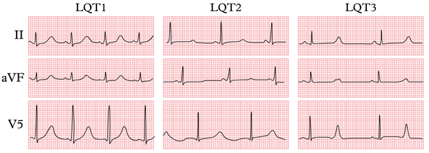
| 18:59, 31 October 2012 | Lqts1-3.png (file) |  |
143 KB | 2 | |
| 21:04, 30 October 2012 | Treatment strategy in HCM.svg (file) |  |
552 KB | 1 | |
| 19:39, 28 October 2012 | Cardiologist menopause physician.svg (file) |  |
22 KB | 1 | |
| 18:44, 25 October 2012 | CRT flowchart.svg (file) |  |
55 KB | 2 | |
| 04:55, 24 October 2012 | Acute hf flowchart.svg (file) |  |
61 KB | 2 | |
| 04:45, 24 October 2012 | Pressure volume curve.svg (file) |  |
27 KB | 2 | |
| 19:04, 22 October 2012 | Suspected heart failure.svg (file) |  |
57 KB | 2 | |
| 18:53, 22 October 2012 | Algorithm for the initial evaluation of patients with clinical symptoms of angina.svg (file) |  |
70 KB | 1 | |
| 17:59, 22 October 2012 | Chest pain to NSTEMI STEMI.svg (file) |  |
28 KB | 1 | |
| 18:35, 18 October 2012 | Frank starling.svg (file) |  |
6 KB | 1 | |
| 18:33, 18 October 2012 | Management outline.svg (file) |  |
12 KB | 1 | |
| 18:31, 18 October 2012 | HF prognosis trials.svg (file) |  |
45 KB | 1 | |
| 05:26, 18 October 2012 | Management chronic systolic hf.svg (file) |  |
589 KB | 1 | |
| 20:24, 14 October 2012 | Process of cardiac remodelling.svg (file) |  |
209 KB | 2 | |
| 16:19, 10 October 2012 | Henle loop.svg (file) |  |
672 KB | 1 |