File:Figure 3.jpg

Size of this preview: 797 × 600 pixels. Other resolutions: 2,560 × 1,927 pixels | 3,898 × 2,934 pixels.
Original file (3,898 × 2,934 pixels, file size: 691 KB, MIME type: image/jpeg)
| Description |
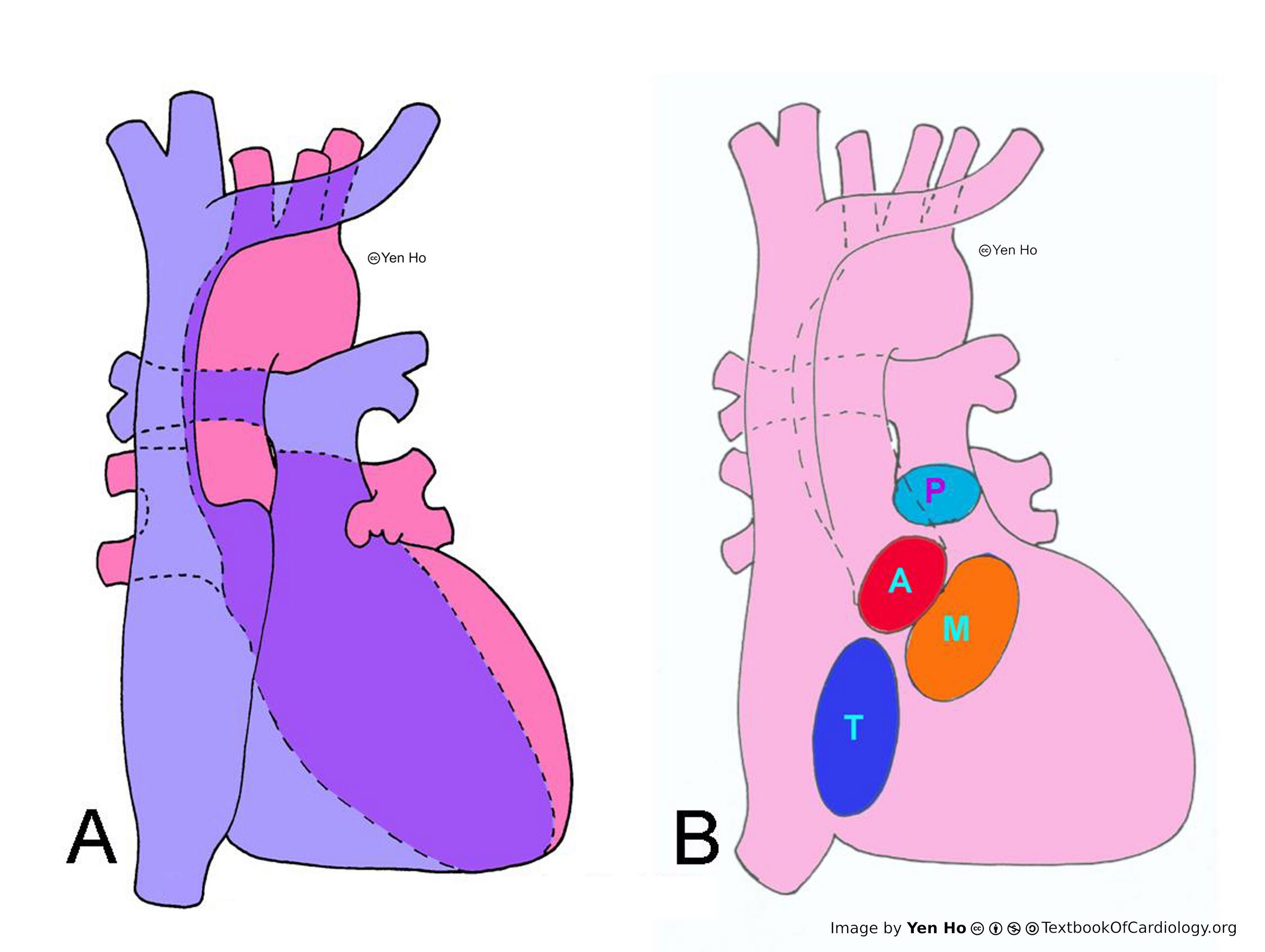
A. Viewed from the front, the right atrium and right ventricle overlaps the left atrium and left ventricle. The atrial chambers are to the right of their respective ventricular chambers.
|
|---|---|
| Source |
provided by S. Yen Ho, PhD FRCPath FESC FHEA, Royal Brompton Hospital, UK |
| Date |
2012 |
| Author |
S. Yen Ho, PhD FRCPath FESC FHEA, Royal Brompton Hospital, UK |
| Permission |
File history
Click on a date/time to view the file as it appeared at that time.
| Date/Time | Thumbnail | Dimensions | User | Comment | |
|---|---|---|---|---|---|
| current | 10:40, 18 May 2012 | | 3,898 × 2,934 (691 KB) | NiloferT (talk | contribs) |
You cannot overwrite this file.
File usage
There are no pages that use this file.