File list
Jump to navigation
Jump to search
This special page shows all uploaded files.
| Date | Name | Thumbnail | Size | User | Description | Versions |
|---|---|---|---|---|---|---|
| 21:50, 31 October 2012 | Arvdhart.png (file) |  |
964 KB | NiloferT (talk | contribs) | 1 | |
| 21:45, 31 October 2012 | Epsilon wave.png (file) |  |
14 KB | NiloferT (talk | contribs) | 1 | |
| 18:59, 31 October 2012 | Lqts1-3.png (file) |  |
143 KB | Drj (talk | contribs) | 2 | |
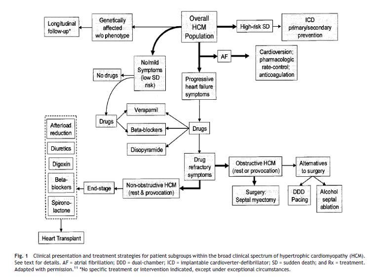
| 21:04, 30 October 2012 | Treatment strategy in HCM.svg (file) |  |
552 KB | Drj (talk | contribs) | 1 | |
| 19:39, 28 October 2012 | Cardiologist menopause physician.svg (file) |  |
22 KB | Drj (talk | contribs) | 1 | |
| 18:44, 25 October 2012 | CRT flowchart.svg (file) |  |
55 KB | Drj (talk | contribs) | 2 | |
| 23:02, 24 October 2012 | Image11.svg (file) |  |
500 KB | April (talk | contribs) | 1 | |
| 23:01, 24 October 2012 | Image9 Takotsubo.svg (file) |  |
147 KB | April (talk | contribs) | 1 | |
| 04:55, 24 October 2012 | Acute hf flowchart.svg (file) |  |
61 KB | Drj (talk | contribs) | 2 | |
| 04:45, 24 October 2012 | Pressure volume curve.svg (file) |  |
27 KB | Drj (talk | contribs) | 2 | |
| 06:39, 23 October 2012 | Test veins.svg (file) |  |
222 KB | April (talk | contribs) | 1 | |
| 19:04, 22 October 2012 | Suspected heart failure.svg (file) |  |
57 KB | Drj (talk | contribs) | 2 | |
| 18:53, 22 October 2012 | Algorithm for the initial evaluation of patients with clinical symptoms of angina.svg (file) |  |
70 KB | Drj (talk | contribs) | 1 | |
| 17:59, 22 October 2012 | Chest pain to NSTEMI STEMI.svg (file) |  |
28 KB | Drj (talk | contribs) | 1 | |
| 18:58, 18 October 2012 | PLAX Mmode.jpg (file) |  |
93 KB | NiloferT (talk | contribs) | Description: Echocardiogram in the parasternal long-axis view, showing a measurement of the heart's left ventricle Date: May, 2005 Author: Ekko (Uploaded Kjetil Lenes, who made the picture. It is released into the public domain.) | 1 |
| 18:50, 18 October 2012 | LeftVentricleShortAxis.gif (file) |  |
64 KB | NiloferT (talk | contribs) | Description: Short axis view of left ventricle of heart Date: 10 July 1999 Source:http://www.yale.edu/imaging/echo_atlas/views/short_axis_lv.html Author: Patrick J. Lynch and C. Carl Jaffe | 1 |
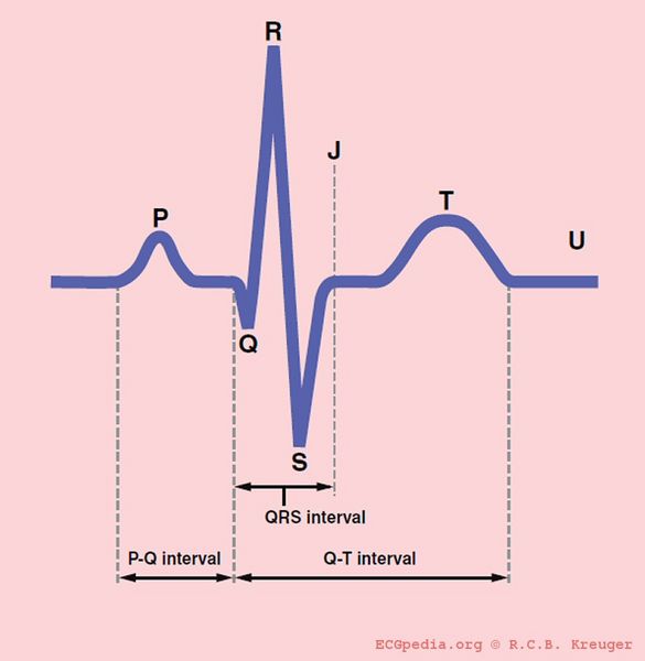
| 18:47, 18 October 2012 | QRSwaves.jpg (file) |  |
20 KB | NiloferT (talk | contribs) | Description: The different waves and intervals of the ECG (P, PQ, QRS, QT, ST) Date: 2007 Author: Rob Kreuger, medical illustrator, AMC, The Netherlands | 1 |
| 18:42, 18 October 2012 | Heart lpla echocardiography diagram.jpg (file) |  |
133 KB | NiloferT (talk | contribs) | Description: Heart normal LPLA left parasternal long axis echocardiography view Date: 23 December 2006 Author: Patrick J. Lynch, medical illustrator | 1 |
| 18:38, 18 October 2012 | Apical 4 chamber view.gif (file) |  |
72 KB | NiloferT (talk | contribs) | Description: Apical four chamber view of heart Date: 10 July 1999 Source: http://www.yale.edu/imaging/echo_atlas/views/four_chamber.html Author: Patrick J. Lynch and C. Carl Jaffe | 1 |
| 18:35, 18 October 2012 | Frank starling.svg (file) |  |
6 KB | Drj (talk | contribs) | 1 | |
| 18:33, 18 October 2012 | Management outline.svg (file) |  |
12 KB | Drj (talk | contribs) | 1 | |
| 18:31, 18 October 2012 | HF prognosis trials.svg (file) |  |
45 KB | Drj (talk | contribs) | 1 | |
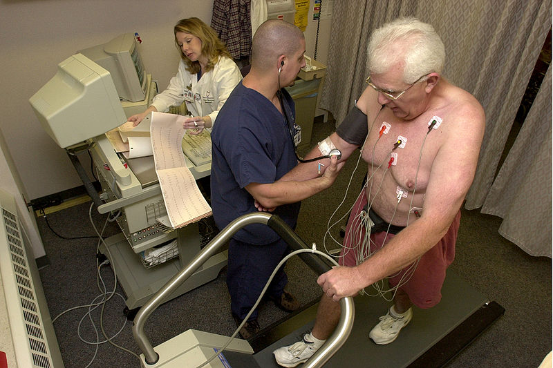
| 18:25, 18 October 2012 | Stress test.jpg (file) |  |
108 KB | NiloferT (talk | contribs) | Description: Stock footage taken at Beaumont Hospital. 14:18, 28 October 2006(UTC) Date: 2006-10-28 Author: Blue0ctane at en.wikipedia | 1 |
| 05:26, 18 October 2012 | Management chronic systolic hf.svg (file) |  |
589 KB | Drj (talk | contribs) | 1 | |
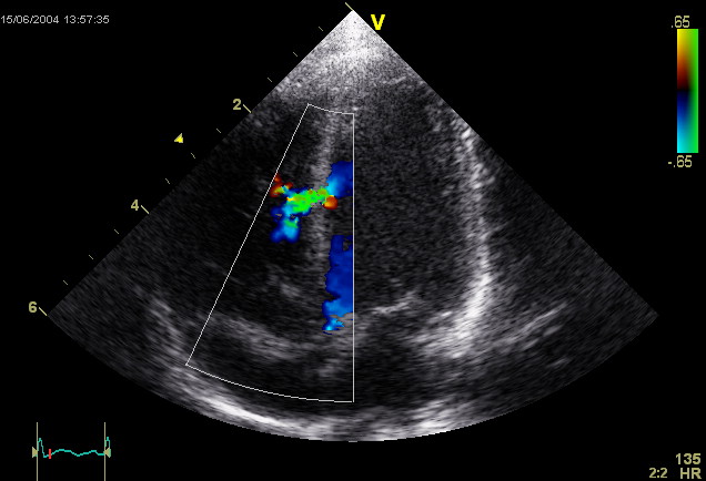
| 23:48, 17 October 2012 | Ventricular Septal Defect.jpg (file) |  |
54 KB | NiloferT (talk | contribs) | This is an ultrasound picture of the heart, an echocardiogram. It depicts a ventricular septal defect. Author: Kjetil Lenes. | 1 |
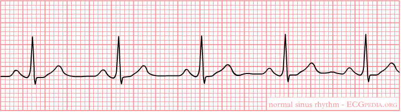
| 23:31, 17 October 2012 | Nsr.png (file) |  |
11 KB | NiloferT (talk | contribs) | 1 | |
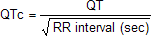
| 23:31, 17 October 2012 | Formule QTc.png (file) | 754 bytes | NiloferT (talk | contribs) | 1 | ||
| 20:24, 14 October 2012 | Process of cardiac remodelling.svg (file) |  |
209 KB | Drj (talk | contribs) | 2 | |
| 16:19, 10 October 2012 | Henle loop.svg (file) |  |
672 KB | Drj (talk | contribs) | 1 | |
| 20:23, 9 October 2012 | Aortic valve (1).gif (file) | .gif) |
2.23 MB | NiloferT (talk | contribs) | This is a video clip from a living, beating pig heart that was prepared in the laboratory as a working Langendorf preparation. The heart was arrested, connected to the perfusion system and restarted. The working fluid was oxygenated balanced saline sol... | 2 |
| 18:30, 9 October 2012 | Pulmonary valve stenosis.svg (file) |  |
59 KB | NiloferT (talk | contribs) | Description: The diagram shows a healthy heart and one suffering from Pulmonary valve stenosis. Date: 12 June 2006 Author: Mariana Ruiz LadyofHats | 1 |
| 18:29, 9 October 2012 | Heart bicuspid aortic valve.svg (file) |  |
28 KB | NiloferT (talk | contribs) | Description: Heart bicuspid aortic valve anatomy Date: 23 December 2006 Author: Patrick J. Lynch, medical illustrator Creative Commons Attribution 2.5 License 2006 | 1 |
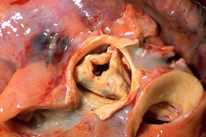
| 18:28, 9 October 2012 | Aortic stenosis rheumatic, gross pathology 20G0014 lores.jpg (file) |  |
92 KB | NiloferT (talk | contribs) | Description: Gross pathology of rheumatic heart disease: aortic stenosis. Aorta has been removed to show thickened, fused aortic valve leaflets and opened coronary arteries from above. Autopsy. Content Providers(s). Author: CDC/Dr. Edwin P. Ewing, Jr. ... | 1 |
| 18:23, 9 October 2012 | Diagram of the human heart (valves improved).svg (file) | .svg) |
27 KB | NiloferT (talk | contribs) | Source:http://commons.wikimedia.org/wiki/Image:Diagram_of_the_human_heart_%28cropped%29.svg | 2 |
| 17:57, 9 October 2012 | LQTS triggers.svg (file) |  |
267 KB | Drj (talk | contribs) | 1 | |
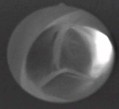
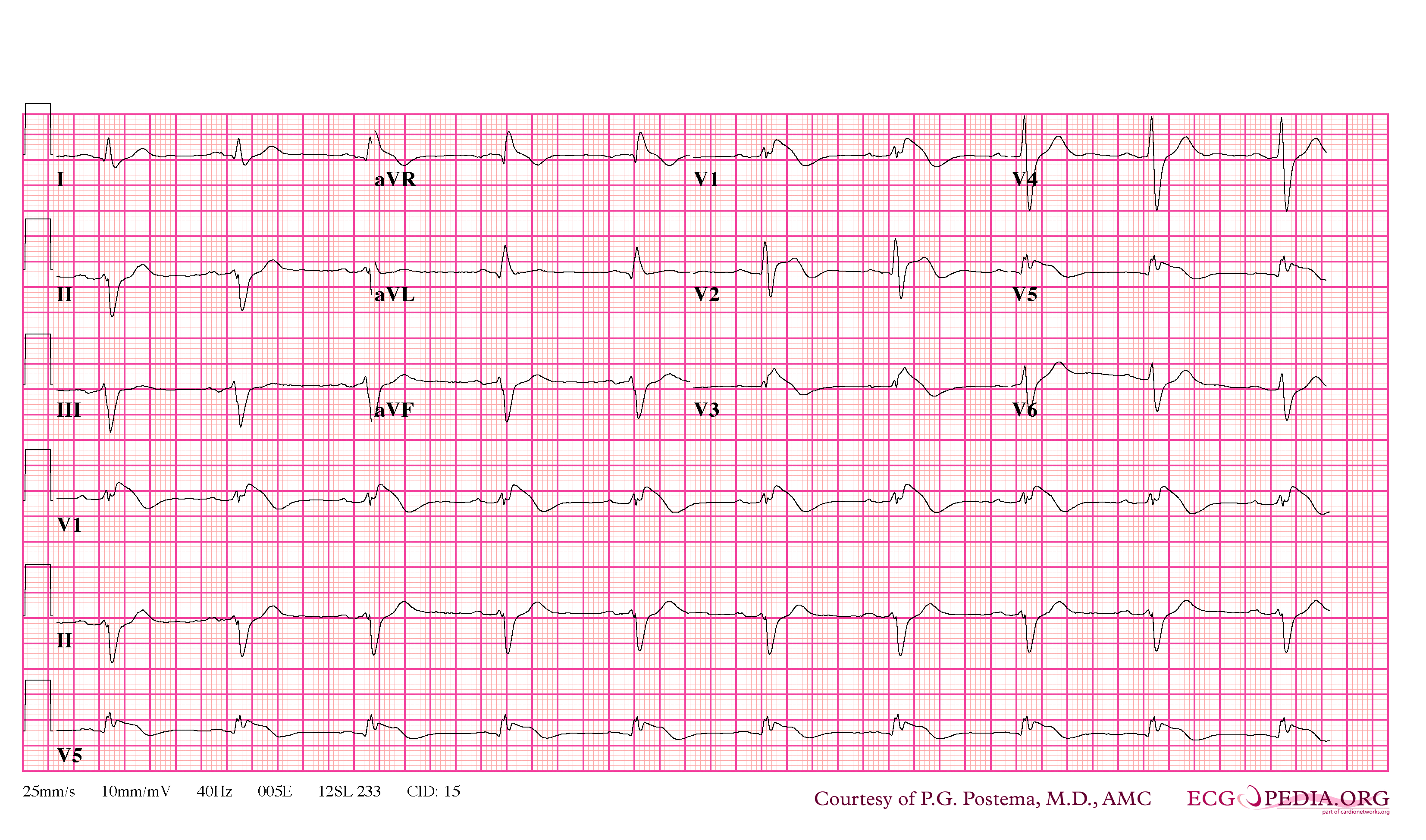
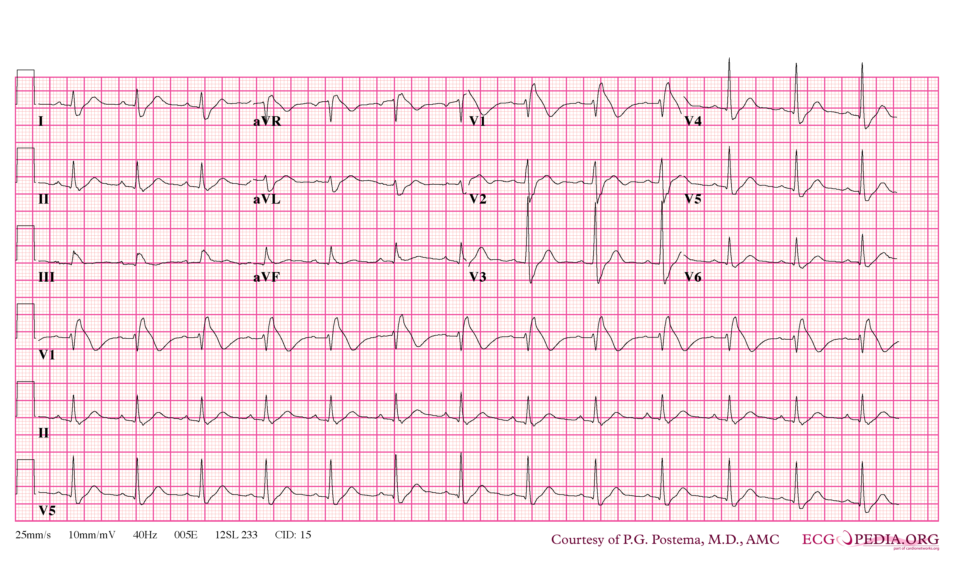
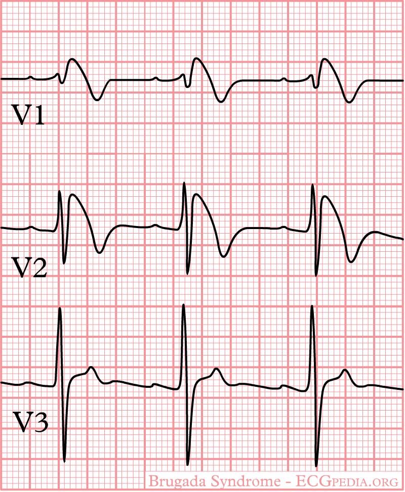
| 09:31, 9 October 2012 | Brugada syndrome type1 example6.jpg (file) |  |
970 KB | NiloferT (talk | contribs) | 1 | |
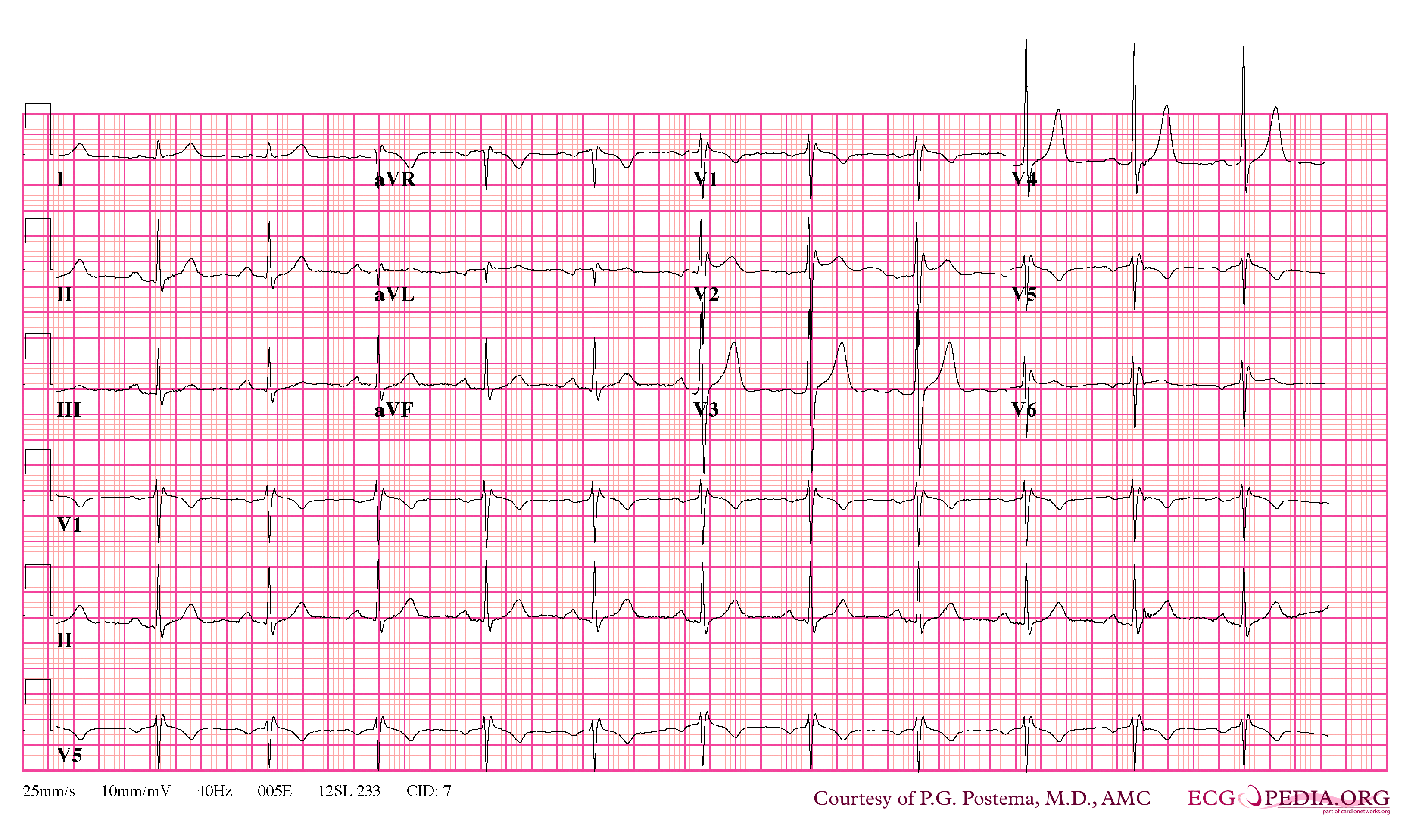
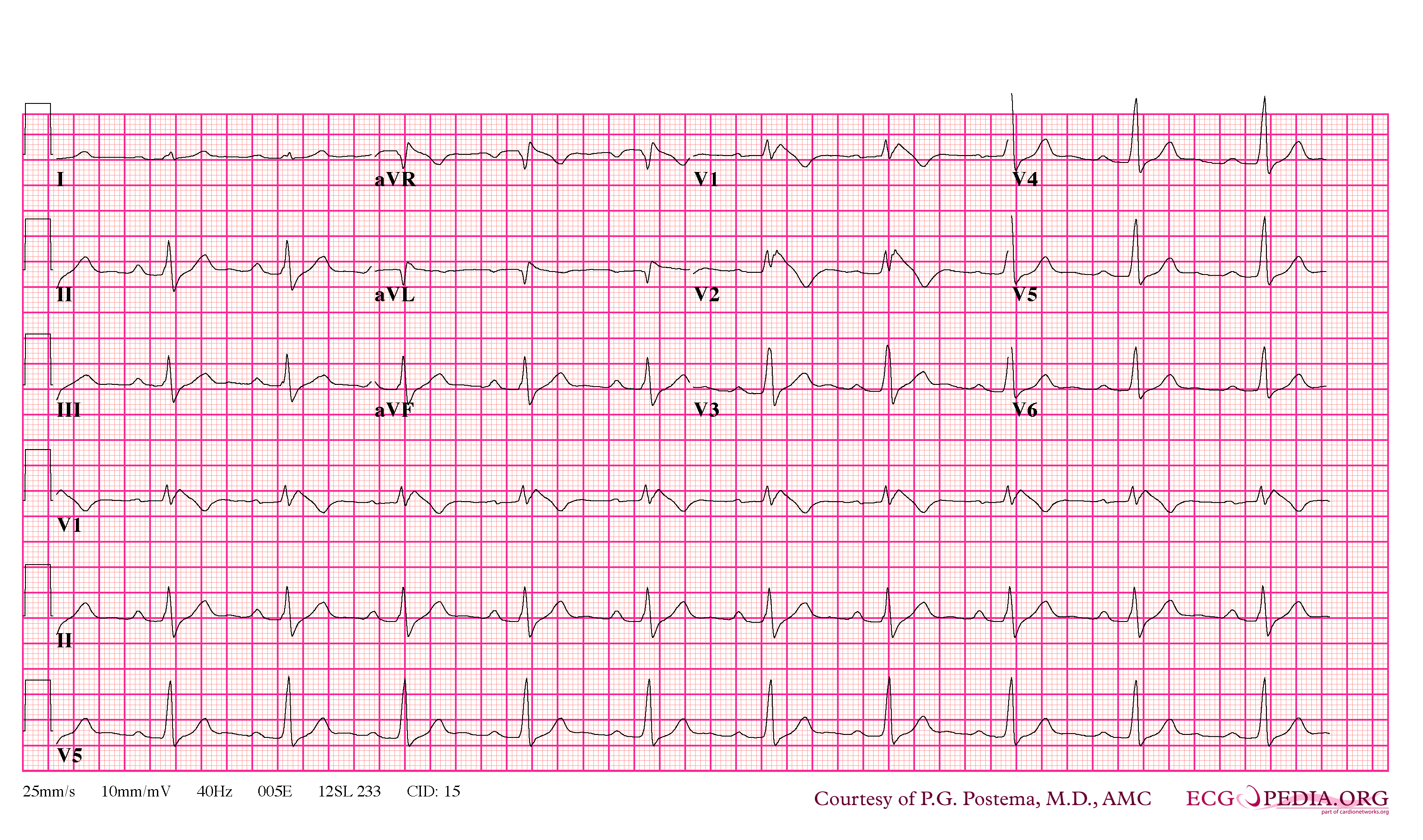
| 09:29, 9 October 2012 | Brugada syndrome type2 example2.jpg (file) |  |
677 KB | NiloferT (talk | contribs) | 1 | |
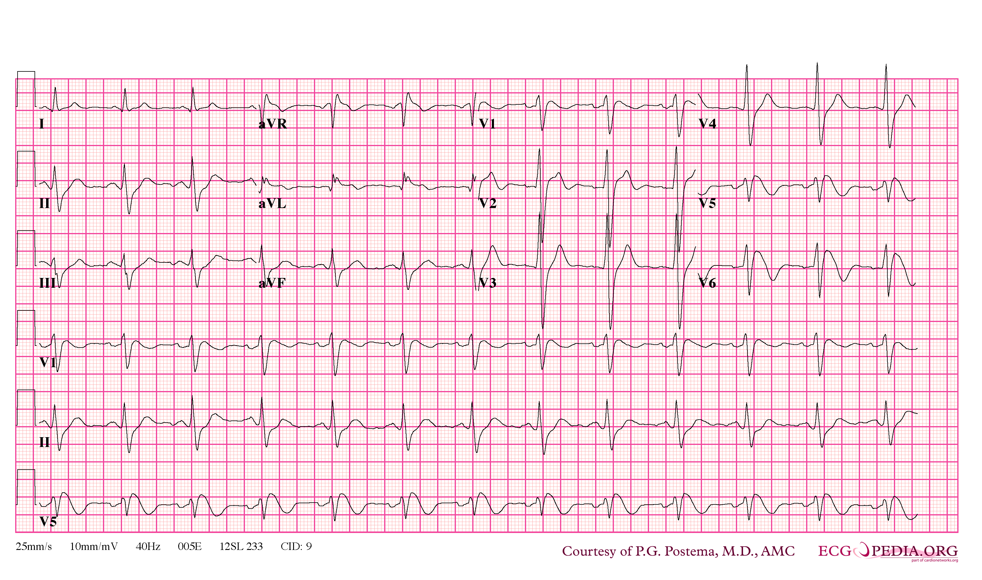
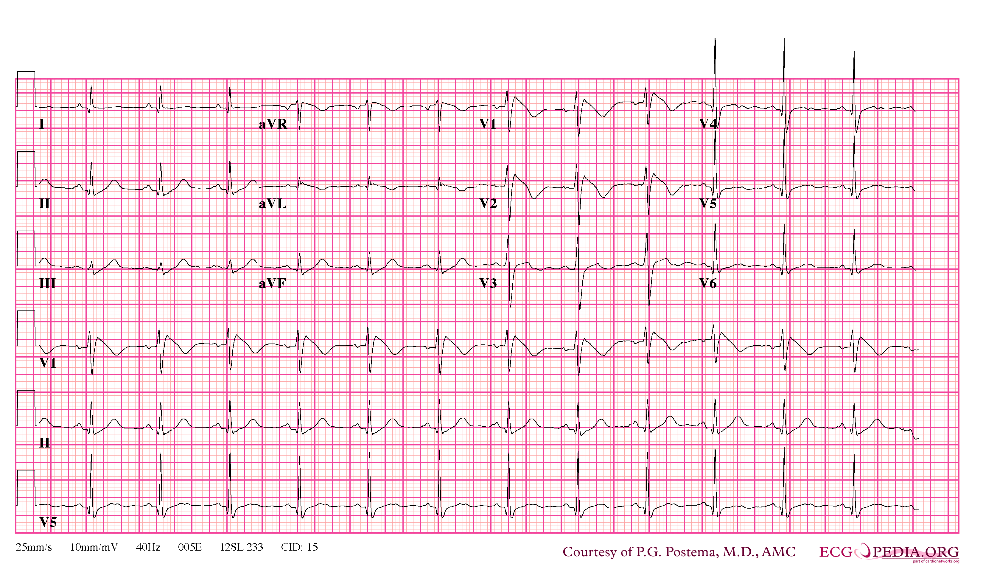
| 09:26, 9 October 2012 | Brugada syndrome type1 example2.png (file) |  |
348 KB | NiloferT (talk | contribs) | 1 | |
| 09:26, 9 October 2012 | Brugada syndrome type2 example1.png (file) |  |
363 KB | NiloferT (talk | contribs) | 1 | |
| 09:25, 9 October 2012 | Brugada syndrome type1 example1.png (file) |  |
370 KB | NiloferT (talk | contribs) | 1 | |
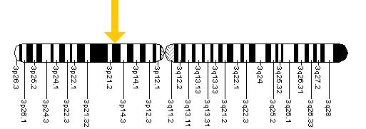
| 09:25, 9 October 2012 | Scn5a.jpg (file) |  |
17 KB | NiloferT (talk | contribs) | 1 | |
| 09:25, 9 October 2012 | Brugada syndrome type1 example5.png (file) |  |
364 KB | NiloferT (talk | contribs) | 1 | |
| 09:24, 9 October 2012 | Brugada syndrome type1 example4.png (file) |  |
364 KB | NiloferT (talk | contribs) | 1 | |
| 09:22, 9 October 2012 | Brugada syndrome type1 example3.png (file) |  |
63 KB | NiloferT (talk | contribs) | 1 | |
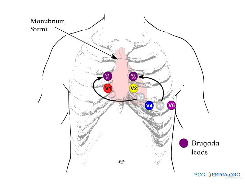
| 09:21, 9 October 2012 | Brugada lead placement.png (file) |  |
84 KB | NiloferT (talk | contribs) | 1 | |
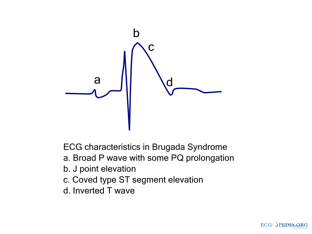
| 09:21, 9 October 2012 | Brugada ecg characteristics.png (file) |  |
39 KB | NiloferT (talk | contribs) | 1 | |
| 09:21, 9 October 2012 | Brugada.png (file) |  |
29 KB | NiloferT (talk | contribs) | 1 | |
| 09:21, 9 October 2012 | Brugada.jpg (file) |  |
13 KB | NiloferT (talk | contribs) | 1 | |
| 17:17, 8 October 2012 | Test.svg (file) |  |
522 KB | Drj (talk | contribs) | 2 | |
| 20:04, 4 October 2012 | Myocardi1.jpg (file) |  |
61 KB | NiloferT (talk | contribs) | 1 |