File list
Jump to navigation
Jump to search
This special page shows all uploaded files.
| Date | Name | Thumbnail | Size | User | Description | Versions |
|---|---|---|---|---|---|---|
| 04:41, 6 August 2012 | AtrialCap.jpg (file) |  |
75 KB | NiloferT (talk | contribs) | 1 | |
| 03:46, 6 August 2012 | ECGT.jpg (file) |  |
57 KB | NiloferT (talk | contribs) | 1 | |
| 03:35, 6 August 2012 | RedTh.jpg (file) | 31 KB | NiloferT (talk | contribs) | 1 | ||
| 02:53, 6 August 2012 | ICD.jpg (file) |  |
67 KB | NiloferT (talk | contribs) | 1 | |
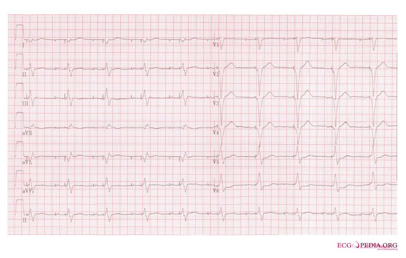
| 00:20, 6 August 2012 | Ddd paced 12lead.jpg (file) |  |
73 KB | NiloferT (talk | contribs) | 1 | |
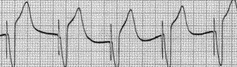
| 00:14, 6 August 2012 | Paced2.gif (file) |  |
7 KB | NiloferT (talk | contribs) | Reverted to version as of 23:31, 5 August 2012 | 3 |
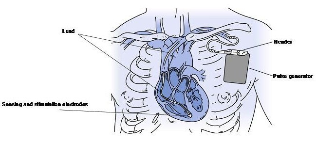
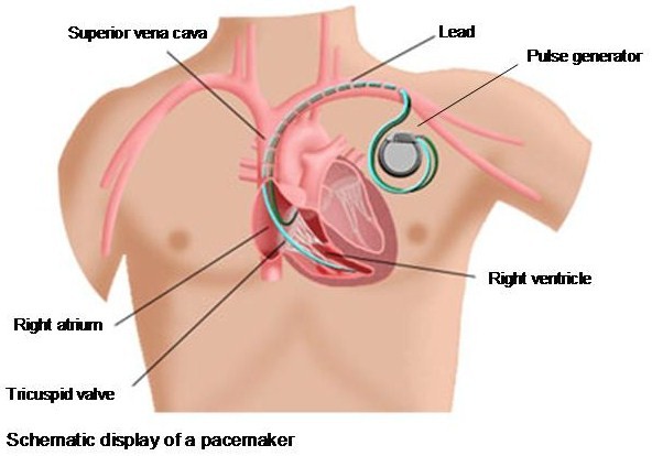
| 22:47, 5 August 2012 | Schematicpic.jpg (file) |  |
53 KB | NiloferT (talk | contribs) | 2 | |
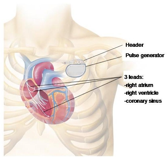
| 22:23, 5 August 2012 | Schematic.jpg (file) |  |
54 KB | NiloferT (talk | contribs) | 1 | |
| 20:14, 5 August 2012 | Xthorax.jpg (file) |  |
44 KB | NiloferT (talk | contribs) | 1 | |
| 19:52, 5 August 2012 | ChestXray.jpg (file) |  |
46 KB | NiloferT (talk | contribs) | 2 | |
| 17:54, 5 August 2012 | SchematicImg.jpg (file) |  |
50 KB | NiloferT (talk | contribs) | 2 | |
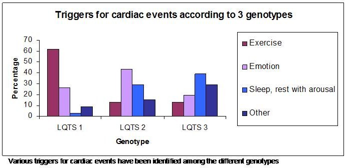
| 02:00, 14 July 2012 | Graph.jpg (file) |  |
52 KB | NiloferT (talk | contribs) | 1 | |
| 20:32, 6 July 2012 | VentDrg.jpg (file) |  |
45 KB | NiloferT (talk | contribs) | 1 | |
| 20:29, 6 July 2012 | TableVent.jpg (file) |  |
126 KB | NiloferT (talk | contribs) | 1 | |
| 19:28, 6 July 2012 | Vent1.jpg (file) |  |
39 KB | NiloferT (talk | contribs) | 1 | |
| 19:19, 6 July 2012 | Bb reentry small.svg (file) | 52 KB | NiloferT (talk | contribs) | 5 | ||
| 04:52, 1 July 2012 | Orthostatic.JPG (file) |  |
25 KB | NiloferT (talk | contribs) | 1 | |
| 04:47, 1 July 2012 | Pathophysiology.JPG (file) |  |
30 KB | NiloferT (talk | contribs) | 1 | |
| 15:25, 20 May 2012 | Figure 7.jpg (file) |  |
1.47 MB | NiloferT (talk | contribs) | Reverted to version as of 15:01, 20 May 2012 | 5 |
| 12:48, 20 May 2012 | Figure 13.jpg (file) |  |
1.48 MB | NiloferT (talk | contribs) | 1 | |
| 12:43, 20 May 2012 | Figure 5.jpg (file) |  |
815 KB | NiloferT (talk | contribs) | 1 | |
| 11:49, 18 May 2012 | Figure 12.jpg (file) |  |
1.99 MB | NiloferT (talk | contribs) | 1 | |
| 11:35, 18 May 2012 | Figure 9.jpg (file) |  |
1.19 MB | NiloferT (talk | contribs) | 2 | |
| 11:30, 18 May 2012 | Figure 11.jpg (file) |  |
1.43 MB | NiloferT (talk | contribs) | 1 | |
| 11:25, 18 May 2012 | Figure 10.jpg (file) |  |
1.3 MB | NiloferT (talk | contribs) | 1 | |
| 11:10, 18 May 2012 | Figure 8.jpg (file) |  |
1.27 MB | NiloferT (talk | contribs) | 1 | |
| 10:50, 18 May 2012 | Figure 6.jpg (file) |  |
601 KB | NiloferT (talk | contribs) | 1 | |
| 10:46, 18 May 2012 | Figure 4.jpg (file) |  |
566 KB | NiloferT (talk | contribs) | 1 | |
| 10:40, 18 May 2012 | Figure 3.jpg (file) |  |
691 KB | NiloferT (talk | contribs) | 1 | |
| 10:36, 18 May 2012 | Figure 2.jpg (file) |  |
1.25 MB | NiloferT (talk | contribs) | 1 | |
| 10:21, 18 May 2012 | Figure1.jpg (file) |  |
1.06 MB | NiloferT (talk | contribs) | 1 | |
| 12:06, 10 May 2012 | AVRT.png (file) |  |
239 KB | Spjkrul (talk | contribs) | 1 | |
| 09:45, 10 May 2012 | AVNRT.png (file) |  |
54 KB | Spjkrul (talk | contribs) | 1 | |
| 09:44, 10 May 2012 | AVNRT.svg (file) |  |
239 KB | Spjkrul (talk | contribs) | 1 | |
| 09:43, 10 May 2012 | Atrial arrhythmias.svg (file) |  |
33 KB | Spjkrul (talk | contribs) | 2 | |
| 18:33, 4 February 2012 | Intraventricular Conduction.svg (file) |  |
431 KB | Drj (talk | contribs) | 2 | |
| 18:30, 4 February 2012 | AVBlock.svg (file) |  |
175 KB | Drj (talk | contribs) | 1 | |
| 18:29, 4 February 2012 | Sinusnode.svg (file) |  |
133 KB | Drj (talk | contribs) | 1 | |
| 18:27, 4 February 2012 | BLS.svg (file) |  |
22 KB | Drj (talk | contribs) | 1 | |
| 16:32, 4 February 2012 | Mechanisms.svg (file) |  |
19 KB | Drj (talk | contribs) | 1 | |
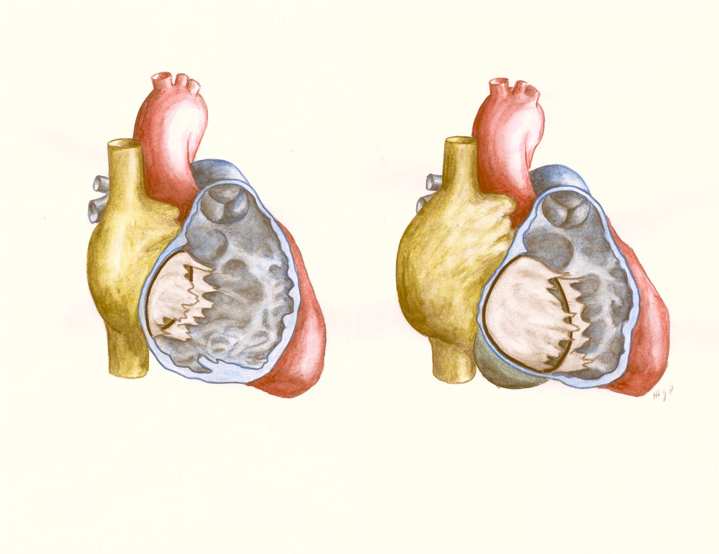
| 16:52, 1 February 2012 | Figure 21. Schematic drawing showing Ebstein’s anomaly of the tricuspid valve.png (file) |  |
1.44 MB | Nja (talk | contribs) | {{Information |Description=Figure 21. Schematic drawing showing Ebstein’s anomaly of the tricuspid valve. Left: normal heart with openend right ventricle. Right: Ebstein’s anomaly with displacement of the septal and posterior tricuspid leaflet, leadin | 1 |
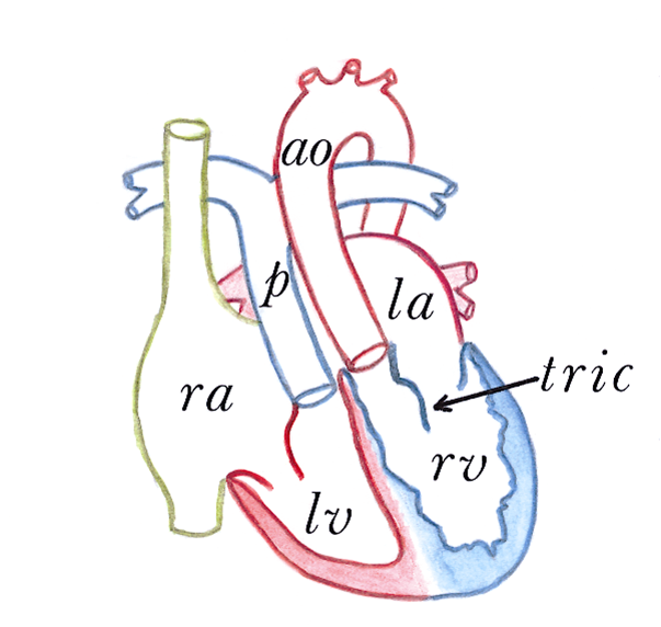
| 16:33, 1 February 2012 | Figure 14. Congenitally corrected transposition of the great arteries.png (file) | 173 KB | Nja (talk | contribs) | {{Information |Description=Figure 14. Congenitally corrected transposition of the great arteries. RA, right atrium. LA, left atrium. RV, right ventricle. LV, left ventricle. p, pulmonary artery. ao, aorta. tric, tricuspid valve. |Source=illustration by dr | 1 | |
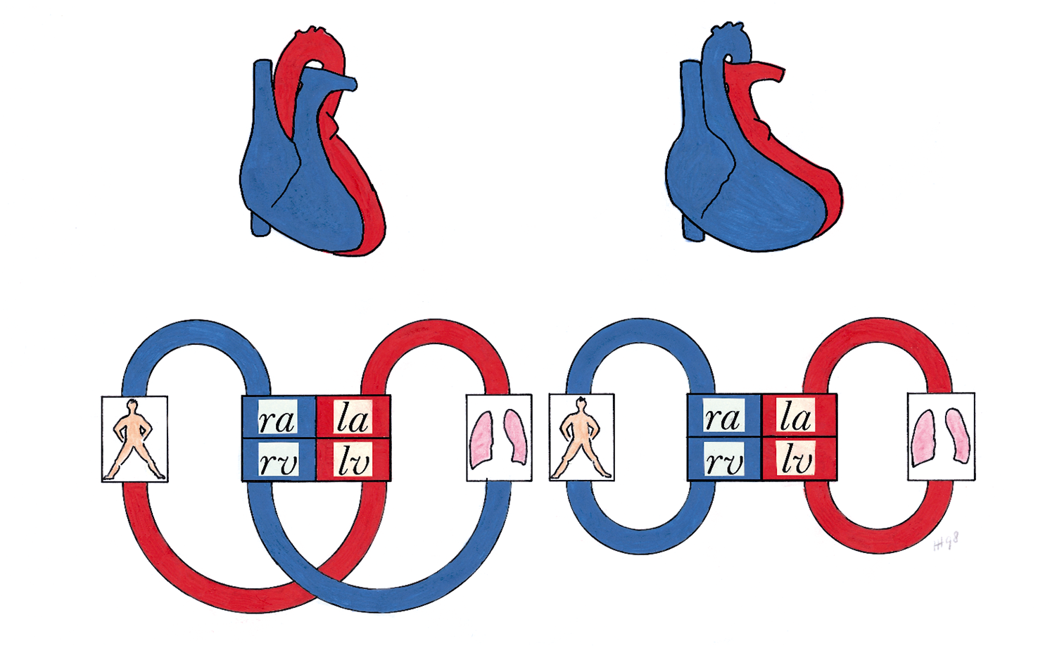
| 16:31, 1 February 2012 | Figure 13. Schematic drawing of the circulation in transposition of the great arteries.png (file) | 821 KB | Nja (talk | contribs) | {{Information |Description=Figure 13. Schematic drawing of the circulation in transposition of the great arteries. Left: normal position of the great arteries with the pulmonary and systemic circulation serially connected. Right: transposition of the grea | 1 | |
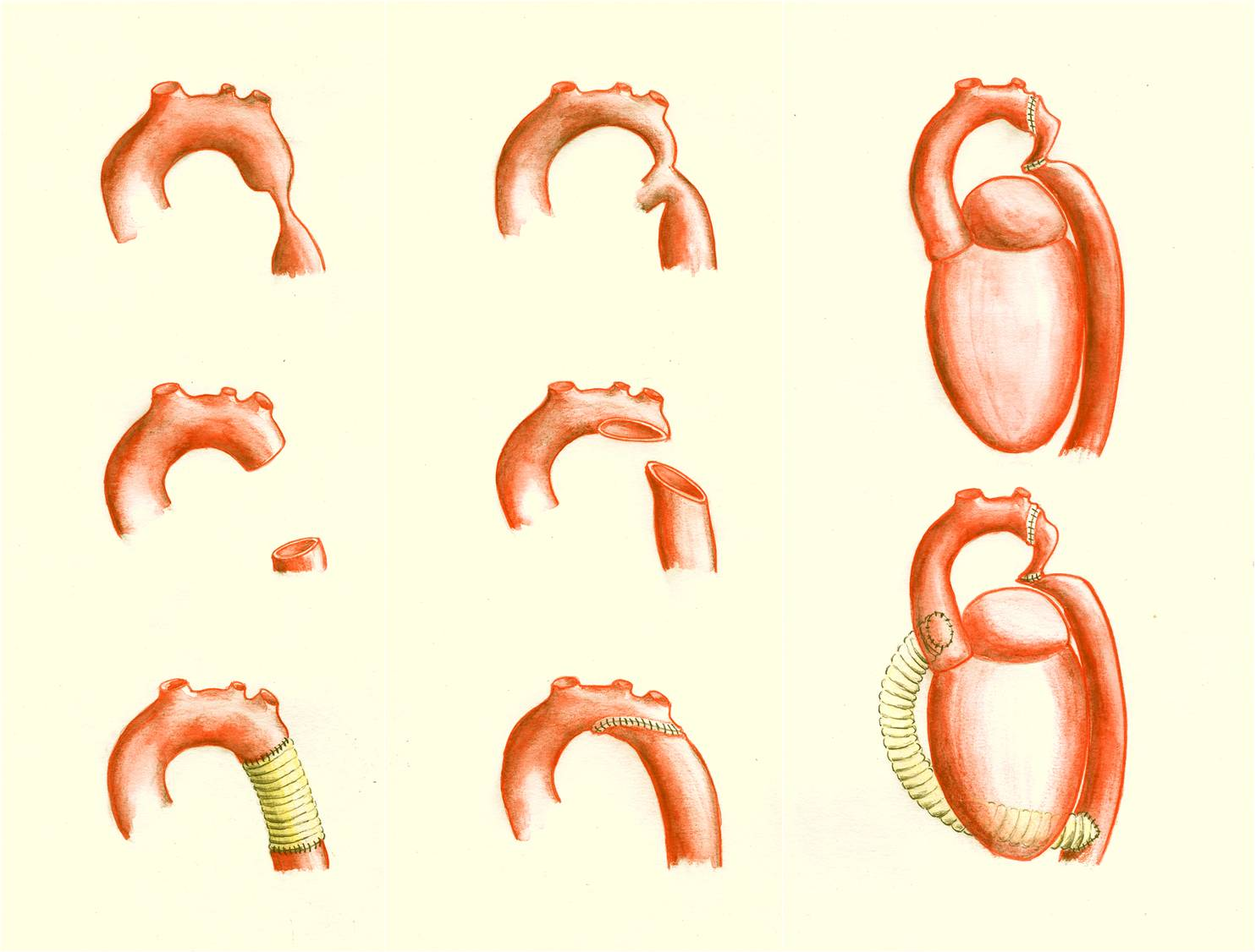
| 16:25, 1 February 2012 | Figure 11. Schematic drawing showing surgical procedures for repair of a coarctation of the aorta.png (file) |  |
1.3 MB | Nja (talk | contribs) | {{Information |Description=Figure 11. Schematic drawing showing surgical procedures for repair of a coarctation of the aorta. Left: an interposition graft. Middle: the extended aortic arch repair. Right: the extra-anatomical bypass. |Source=cillustration | 1 |
| 16:19, 1 February 2012 | Figure 10. Schematic drawing showing surgical procedures for repair of coarctation of the aorta.png (file) |  |
1.22 MB | Nja (talk | contribs) | {{Information |Description=Figure 10. Schematic drawing showing surgical procedures for repair of coarctation of the aorta. Left: resection with end-to-end anastomosis. Middle: dilating technique using a patch; this technique is used in coarctations invol | 1 |
| 16:10, 1 February 2012 | Figure 9. Schematic drawing of the anatomy prenatal and postnatal.png (file) |  |
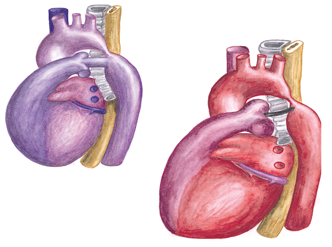
1.24 MB | Nja (talk | contribs) | {{Information |Description=Figure 9. Schematic drawing of the anatomy prenatal (left) and postnatal (right) in coarctation of the aorta. In the normal situation (without coarctation) only 10 percent of the fetal cardiac output flows through the descending | 1 |
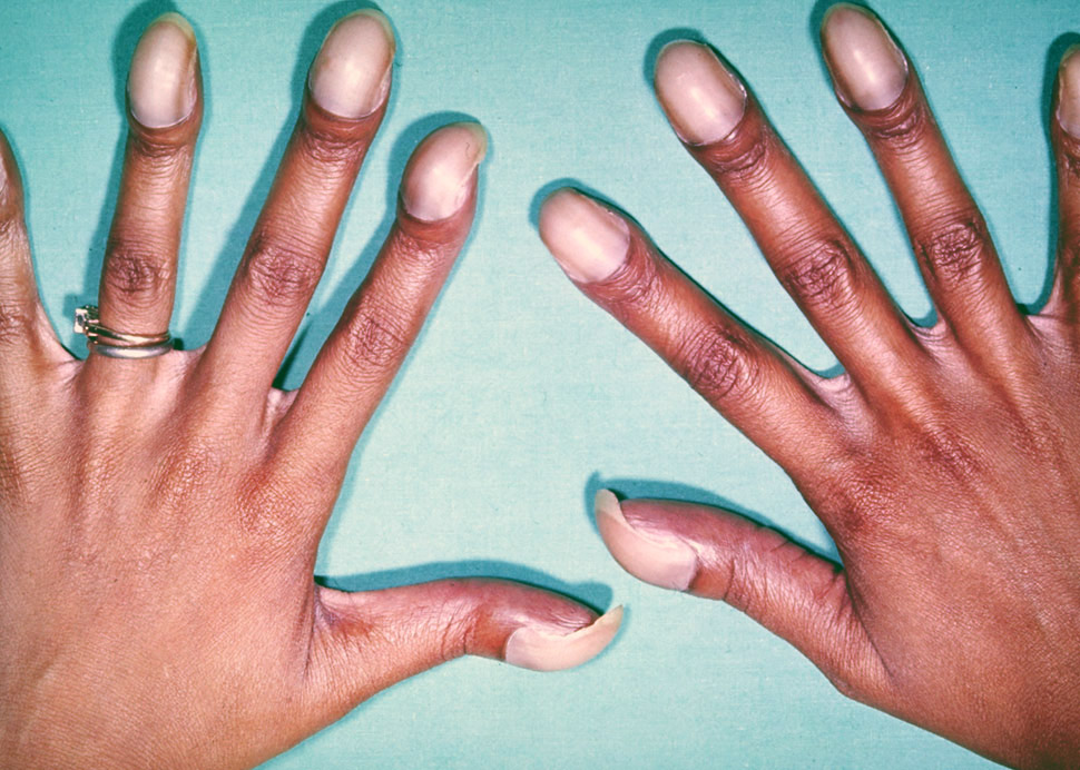
| 15:22, 25 January 2012 | 22. Eisenmenger.jpg (file) |  |
219 KB | Nja (talk | contribs) | {{Information |Description=Figure 22. Photo showing typical features of chronic hypoxemia in Eisenmenger syndrome, with typical digital clubbing with cyanotic nail beds. |Source=from commons.wikipedia.org |Date=Published: |Author= |Permission= |other_vers | 1 |
| 15:19, 25 January 2012 | 21. Ebstein.PNG (file) |  |
495 KB | Nja (talk | contribs) | {{Information |Description=Figure 21. Schematic drawing showing Ebstein’s anomaly of the tricuspid valve. Left: normal heart with openend right ventricle. Right: Ebstein’s anomaly with displacement of the septal and posterior tricuspid leaflet, leadin | 1 |
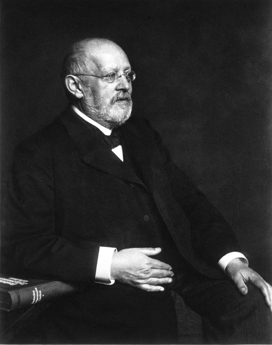
| 15:17, 25 January 2012 | 20. Wilhelm Ebstein.jpg (file) |  |
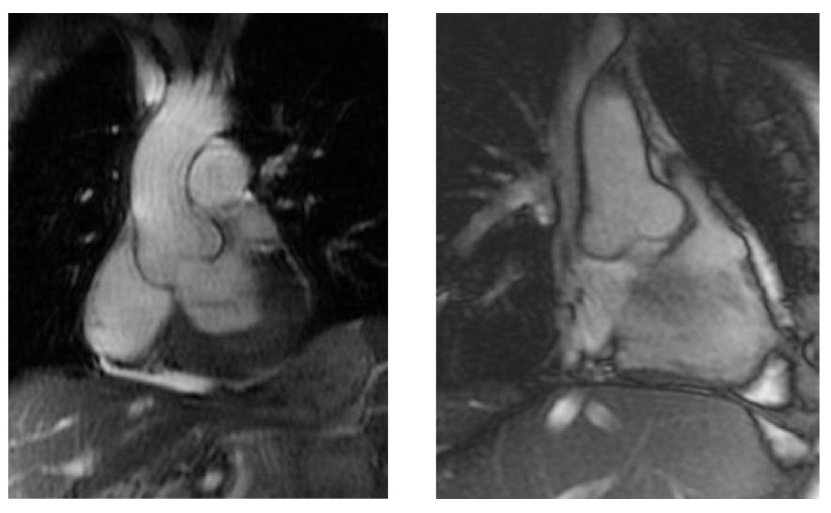
134 KB | Nja (talk | contribs) | {{Information |Description=Figure 19. Magnetic resonance imaging of the aorta, showing aortic root dilatation in Marfan syndrome. |Source=from commons.wikipedia.org |Date=Published: |Author= |Permission= |other_versions= }} | 1 |
| 15:15, 25 January 2012 | 19. MFS2.jpg (file) |  |
89 KB | Nja (talk | contribs) | {{Information |Description=Figure 19. Magnetic resonance imaging of the aorta, showing aortic root dilatation in Marfan syndrome. |Source=from commons.wikipedia.org |Date=Published: |Author= |Permission= |other_versions= }} | 1 |