File:7. PDA.png
No higher resolution available.
7._PDA.png (636 × 432 pixels, file size: 106 KB, MIME type: image/png)
| Description |
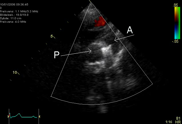
Figure 7. Echocardiographic image showing a coil in the ductus arteriosus. P=pulmonary artery, A= aorta. |
|---|---|
| Source |
from commons.wikipedia.org |
| Date |
Published: |
| Author | |
| Permission |
File history
Click on a date/time to view the file as it appeared at that time.
| Date/Time | Thumbnail | Dimensions | User | Comment | |
|---|---|---|---|---|---|
| current | 14:18, 25 January 2012 | | 636 × 432 (106 KB) | Nja (talk | contribs) | {{Information |Description=Figure 7. Echocardiographic image showing a coil in the ductus arteriosus. P=pulmonary artery, A= aorta. |Source=from commons.wikipedia.org |Date=Published: |Author= |Permission= |other_versions= }} |
You cannot overwrite this file.
File usage
The following page uses this file: