Myocardial Infarction
In 2006 425.425 people died from a heart attack, 1.255.000 new and recurrent coronary attacks took place, about 34% died, 17.600.000 victims of angina, heart attack and other forms of coronary heart disease are still living.
These numbers only account for the United States.
Pathofysiology
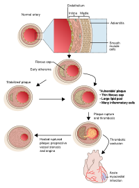
A heart attack or myocardial infarction (MI) is an acute presentation of a process that has been going on much longer. The process responsible is atherosclerosis. Atherosclerosis is a chronic disease of the arteries in which artery walls thicken by deposition of fatty materials such as cholesterol. The result over decades are plaques, which can narrow the lumen of the arteries significantly and progressively causing symptoms as angina pectoris. Plaques can also suddenly rupture, trigger a cascade which results in a thrombus and thereby cause myocardial infarction.
History
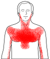
Classic presentation of a myocardial infarction is acute chest pain which lasts longer than a few minutes. The pain does not decrease at rest and is only temporarily relieved with nitroglycerin. Common accompanying symptoms are radiating pain to shoulder, arm, back and/or jaw. Shortness of breath can occur, as well as sweating, fainting, nausea and vomiting, so called vegetative symptoms. Some patients not really complain about chest pain but more about abdominal pain so as with angina pectoris the presentation can be very a specific.
It is important to complete the history with information about past history (prior history of ischemic events or vascular disease), risk factors for cardiovascular disease (o.a. smoking, hypertension, hyperlipidemia, obesity) and family history (direct family with myocardial infarction and/or sudden cardiac death).
Signs of heart failure such as orthopnea (not able to sleep without a pillow), progressive dyspnoea and oedematous ankles are indicative for the extent of the problem.
A suspected myocardial infarction should be rapidly evaluated to initiate appropriate therapy.
Physical Examination
On physical examination evidence of systemic hypoperfusion can be found such as hypotension, tachycardia, impaired cognition, pale and ashen skin. If during auscultation pulmonary crackles are heard and pitting oedema of the ankles is seen heart failure is complicating the myocardial infarction.
History and physical examination are helpful to determine myocardial infarction as diagnosis and to exclude other causes of chest pain, such as angina pectoris, aorta dissection, arrhythmias, pulmonary embolism, pneumonia, heartburn, hyperventilation or musculoskeletal problems.
Electrocardiogram
An electrocardiogram (ECG) should be made within 10 minutes of arrival in every patient with suspected myocardial infarction.
An ECG is important to differentiate between myocardial ischemia and infarction:
- ST elevation in myocardial infarction
- ST depression in myocardial ischemia
And to differentiate between STEMI and NSTEMI:
- STEMI stands for ST elevated (>20 min) Myocardial Infarction
- NSTEMI stand for Non ST elevated Myocardial Infarction
It can however take 90 minutes after the onset of the symptoms to see abnormalities on the ECG. Therefore it is important to make a serial ECG, certainly if a patient has ongoing symptoms.
An ECG is also helpful in localising the ischemia: Anterior wall ischemia - One or more of leads V1-V6 Anteroseptal ischemia - Leads V1 to V3 Apical or lateral ischemia - Leads aVL and I, and leads V4 to V6 Inferior wall ischemia - Leads II, III, and aVF
Cardiac Markers
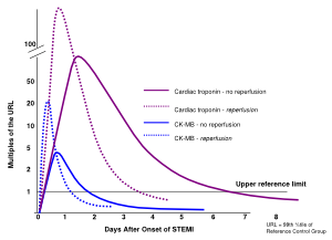
Cardiac markers are essential for confirming the diagnosis of infarction. Elevated CK MB and Troponin I indicate damage of the myocardium. It can however take 4-8 hours, after the symptoms started, before the cardiac markers are elevated. The advise is to repeat the measurements after 4-6 hours. A pitfall concerning elevated Troponin I can be patients with renal failure or pulmonary embolism. Although cardiac markers are helpful for confirming the diagnosis reperfusion should not always wait till the cardiac markers are known.
Treatment
ST elevated Myocardial Infarct
Initial treatment of STEMI is relief of ischemic pain, stabilize the hemodynamic status and reduce the ischemia as quickly as possible by fibrinolysis or primary percutaneous coronary intervention (PCI). Meanwhile other measures as continuous cardiac monitoring, oxygen and intravenous access are necessary to guarantee the safety of the patient.
Rapid revascularisation is essential to minimize the impact of the myocardial infarction and thereby reduce mortality. In the first hours after symptom onset the amount of salvageable myocardium by reperfusion is greatest. Revascularisation can be achieved by fibrinolysis or PCI.
PCI is, if available, the preferred revascularisation method for patients with STEMI. But not all hospitals are qualified to perform PCI and therefore fibrinolysis is still used. There are however some circumstances in which transfer to a PCI qualified hospital is essential:
- Patients with contraindications for fibrinolysis as active bleedings, recent dental surgery, past history of intracranial bleeding.
- Patients with cardiogenic shock, severe heart failure and/or pulmonary oedema complicating the myocardial infarction.
Or when PCI has a better outcome:
- Patients who present three hours to four hours after the onset of the symptoms.
- Patients with a non diagnostic ECG or a atypical history a coronary angiography with the ability to perform a PCI is preferred.
Fibrinolysis
Fibrinolytics like streptokinase stimulate the conversion of plasminogen to plasmin. Plasmin demolishes fibrin which is an important constituent of the thrombus. Fibrinolytics are most effective the first hours after the onset of symptoms, after twelve hours the outcome will not improve. Because re occlusion after fibrinolysis is possible patients should be transferred to a PCI qualified hospital once fibrinolysis is done.
Percutaneous Coronary Intervention (PCI)
| align=right|height=300px|width=300px</flashow> | align=right|height=300px|width=300px</flashow> |
| Right coronary artery (RCA) occlusion before PCI | Reflow in the RCA after PCI |
The procedure of PCI starts off as a coronary angiography (see CAG). When the stenosis is visualized a catheter with an inflatable balloon will be brought at the site of the stenosis. Inflation of the balloon within the coronary artery will crush the atherosclerosis and eliminate the stenosis. To prevent that the effect of the balloon is only temporarily a stent is positioned at the site of the stenosis. To reduce the risk of coronary artery stent thrombosis antiplatelet therapy should be given.
Coronary Artery Bypass Graft
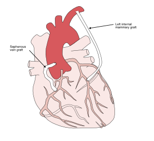
When the coronary arteries contain too many or too severe stenoses for PCI a coronary artery bypass graft (CABG) is indicated. Especially when the stenoses are located proximally of the three major coronary arteries, causing occlusion of many ramifications and high risk of severe myocardial damage.
CABG does not eliminate the stenosis like PCI does. Using the internal thoracic arteries or the saphenous veins from the legs a bypass is made around the stenosis. The bypass originates from the aorta and terminates directly after the stenosis. Thereby restoring the blood supply to the ramifications. A bypass can be single or multiple, multiple meaning that there are several coronary arteries bypassed using the same bypass.
Major surgery is not preferable in patients with STEMI, but CABG is inevitable when fibrinolysis and/or PCI failed or when the patient develops cardiogenic shock, life threatening ventricular arrhymthmias or three vessel disease.
Medication to start after MI
β blockers lower heart rate and blood pressure, this decreases the oxygen demand of the heart. Nitrates dilatate the coronary arteries so the heart receives more oxygenated blood. Anticoagulants reduce the risk of development of a thrombus in the coronary arteries. Statins: Apart from starting medication the patient needs to minimize any present risk factors like smoking, overweight and drinking alcohol.
Non ST elevated Myocardial Infarct
Initial treatment in NSTEMI is to reduce ischemia, stabilize the hemodynamic status, make serial ECG and to repeat measurements of the cardiac markers. Depending on the early risk stratification a choice has to be made between early invasive therapy or conservative therapy with medicines.
Early risk stratification is helpful to identify patients at high risk who need a more aggressive therapeutic approach to prevent further ischemic events.
- Age ≥65 years
- Presence of at least three risk factors for coronary heart disease (hypertension, diabetes, dyslipidemia, smoking, or positive family history of early MI)
- Prior coronary stenosis of ≥50 percent
- Presence of ST segment deviation on admission ECG
- At least two anginal episodes in prior 24 hours
- Elevated serum cardiac biomarkers
Patients with a score of 0 to 1 are at low risk, score 2 to 3 are at intermediate risk, score 4 to 6 are at high risk.
Conservative Therapy
The main objective of in hospital conservative therapy is to relieve ischemic pain by intensifying medical therapy with aspirin and clopidogrel orally and nitro-glycerine, heparin and a beta blocker intravenously. If the patients becomes asymptomatic on these medication and is still asymptomatic when the medication is stopped, rest and stress imaging testing will be performed. The advantage of conservative therapy is reduction of the number of unnecessary revascularizations. The disadvantage is a prolonged stay in the hospital.
Rest and Stress Imaging Tests
Rest and stress testing is indicated in patients with:
- Angina pectoris with ECG abnormalities during exercise ECG testing
- Asymptomatic NSTEMI after in hospital conservative therapy
Exercise echocardiography means that an echocardiography is made directly after exercise. The poorly perfused parts of the heart will show less activity.
Myocardium Perfusion Scintigraphy (MPS) is able to show the perfusion of the heart during exercise and at rest.
MRI can be done with vasodilatory dobutamine or stimulating adenosine to assess how the heart behaves during exercise.
Invasive Therapy
High risk patients, patients with persistent symptoms despite medication or a positive stress test need invasive therapy. Depending on what is seen during coronary angiography PCI or a CABG is indicated. (see PCI/CABG)
Fibrinolytic therapy is not used in NSTEMI .


