File list
Jump to navigation
Jump to search
This special page shows all uploaded files.
| Date | Name | Thumbnail | Size | User | Description | Versions |
|---|---|---|---|---|---|---|
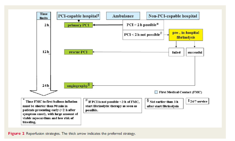
| 14:12, 13 January 2012 | Figure 2 - Reperfusion strategies.png (file) |  |
123 KB | Nja (talk | contribs) | Reperfusion strategies. The thick arrow indicates the preferred strategy. | 1 |
| 12:47, 13 January 2012 | Figure 1 - algorithm for the initial evaluation of patients with clinical symptoms of angina.png (file) |  |
149 KB | Nja (talk | contribs) | Figure 1 Algorithm for the initial evaluation of patients with clinical symptoms of angina | 1 |
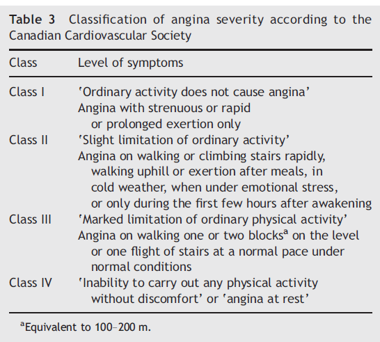
| 12:46, 13 January 2012 | Table 3 - classification of angina severity according to the Canadian Cardiovascular Society.png (file) |  |
77 KB | Nja (talk | contribs) | Classification of Angina Severity According to the Canadian Cardiovascular Society | 1 |
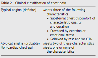
| 12:45, 13 January 2012 | Table 2 - classification of chest pain.png (file) |  |
16 KB | Nja (talk | contribs) | Classification of Chest Pain | 1 |
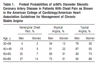
| 12:45, 13 January 2012 | Table 1 - pretest probabilities.png (file) |  |
18 KB | Nja (talk | contribs) | Pretest probabilities of >= 50% Diameter Stenotic Coronary Artery Disease in Patients with Chest Pain | 1 |
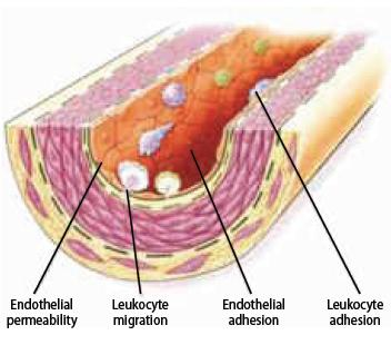
| 01:11, 11 January 2012 | Figure 8 - Endothelial dysfunction - Leukocyte adhesion and migration into the deep layer of the intima.png (file) |  |
188 KB | Nja (talk | contribs) | Figure 8. Endothelial dysfunction: Leukocyte adhesion and migration into the deep layer of the intima. | 1 |
| 01:06, 11 January 2012 | Figure 7 - Fatty streak formation revealing platelet aggregation on the endothelial surface.png (file) |  |
138 KB | Nja (talk | contribs) | Figure 7. Endothelial dysfunction: Leukocyte adhesion and migration into the deep layer of the intima. | 1 |
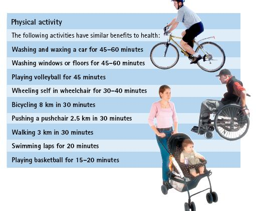
| 13:03, 10 January 2012 | Figure 14 - Recommendations for physical activity.png (file) |  |
212 KB | Nja (talk | contribs) | Figure 14. Recommendations for physical activity | 1 |
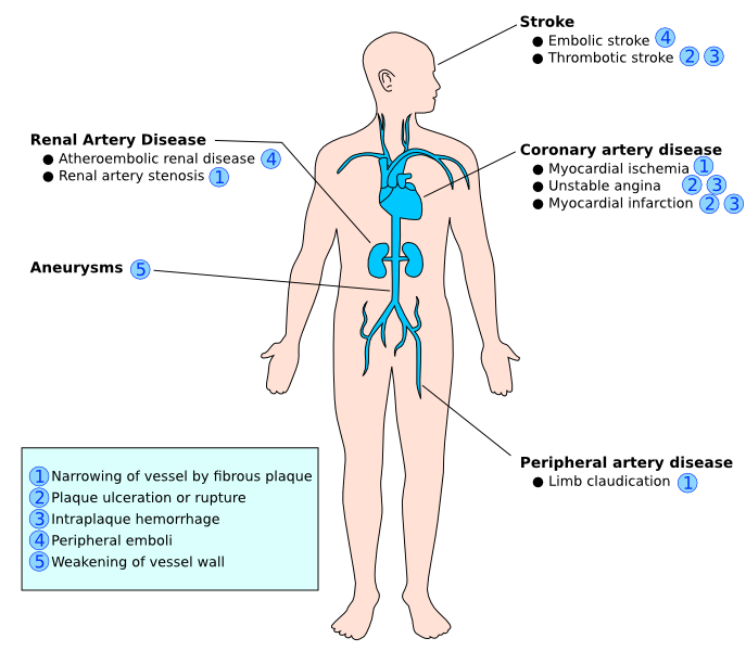
| 13:02, 10 January 2012 | Figure 11 - Complications of atherosclerosis.png (file) |  |
99 KB | Nja (talk | contribs) | Figure 11. Complications of atherosclerosis | 1 |
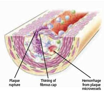
| 13:01, 10 January 2012 | Figure 10 - The ruptured plaque..png (file) |  |
126 KB | Nja (talk | contribs) | Figure 10. The ruptured plaque | 1 |
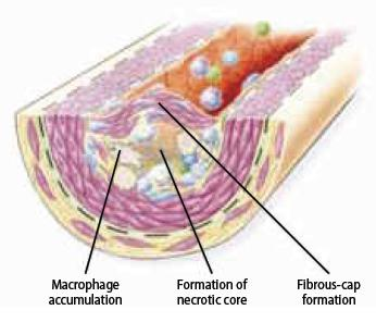
| 13:00, 10 January 2012 | Figure 9 - Fibrous cap formation.png (file) |  |
123 KB | Nja (talk | contribs) | Figure 9. Fibrous cap formation | 1 |
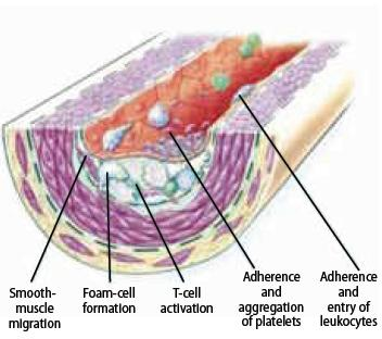
| 13:00, 10 January 2012 | Figure 8 - Fatty streak formation.png (file) |  |
138 KB | Nja (talk | contribs) | Figure 8. Fatty streak formation | 1 |
| 12:52, 10 January 2012 | Figure 7 - Endothelial dysfunction.png (file) |  |
142 KB | Nja (talk | contribs) | Figure 7. Endothelial dysfunction | 1 |
| 12:49, 10 January 2012 | Figure 4 - Distribution of CVD death among females in 2008.png (file) |  |
80 KB | Nja (talk | contribs) | Figure 4. Distribution of CVD death among females in 2008 | 1 |
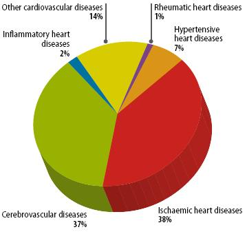
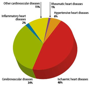
| 12:46, 10 January 2012 | Figure 3 - Distribution of CVD death among males in 2008.png (file) |  |
77 KB | Nja (talk | contribs) | Figure 3. Distribution of CVD death among males in 2008 | 1 |
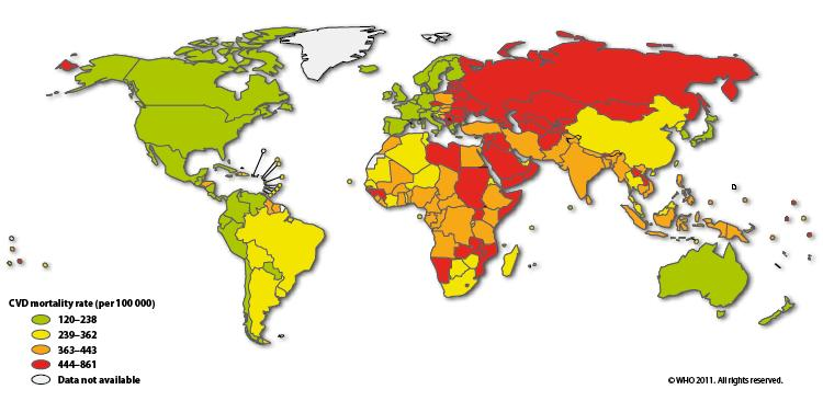
| 12:45, 10 January 2012 | Figure 2 - World map CVD mortality rates in females.png (file) |  |
249 KB | Nja (talk | contribs) | Figure 2. World map CVD mortality rates in females | 1 |
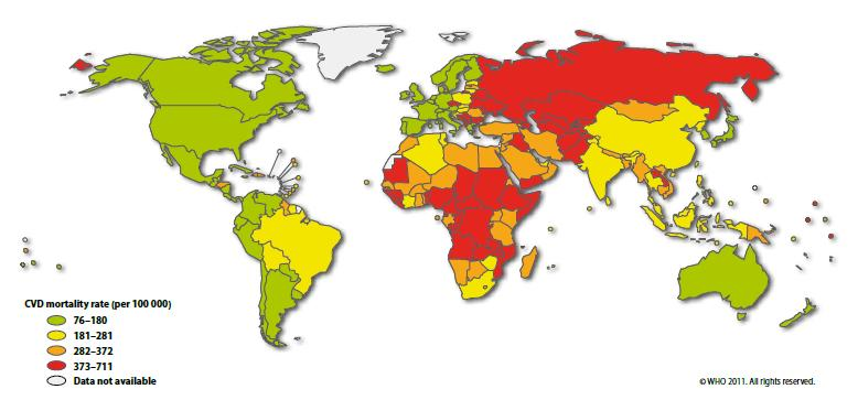
| 12:44, 10 January 2012 | Figure 1 - World map CVD mortality rates in males.png (file) |  |
240 KB | Nja (talk | contribs) | Figure 1. World map CVD mortality rates in males | 1 |
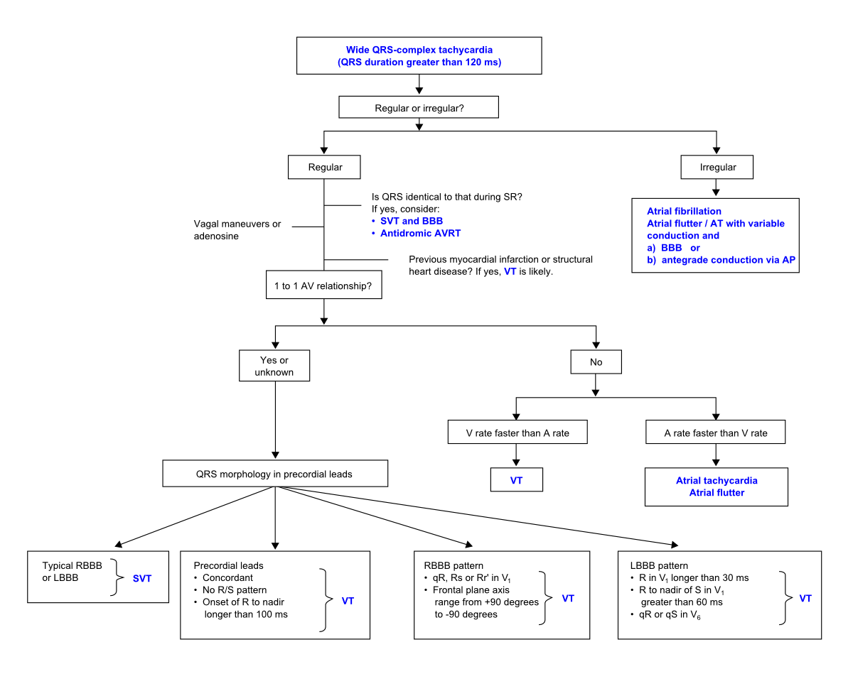
| 16:44, 28 December 2011 | BroadTachydiff.png (file) |  |
90 KB | Spjkrul (talk | contribs) | 2 | |
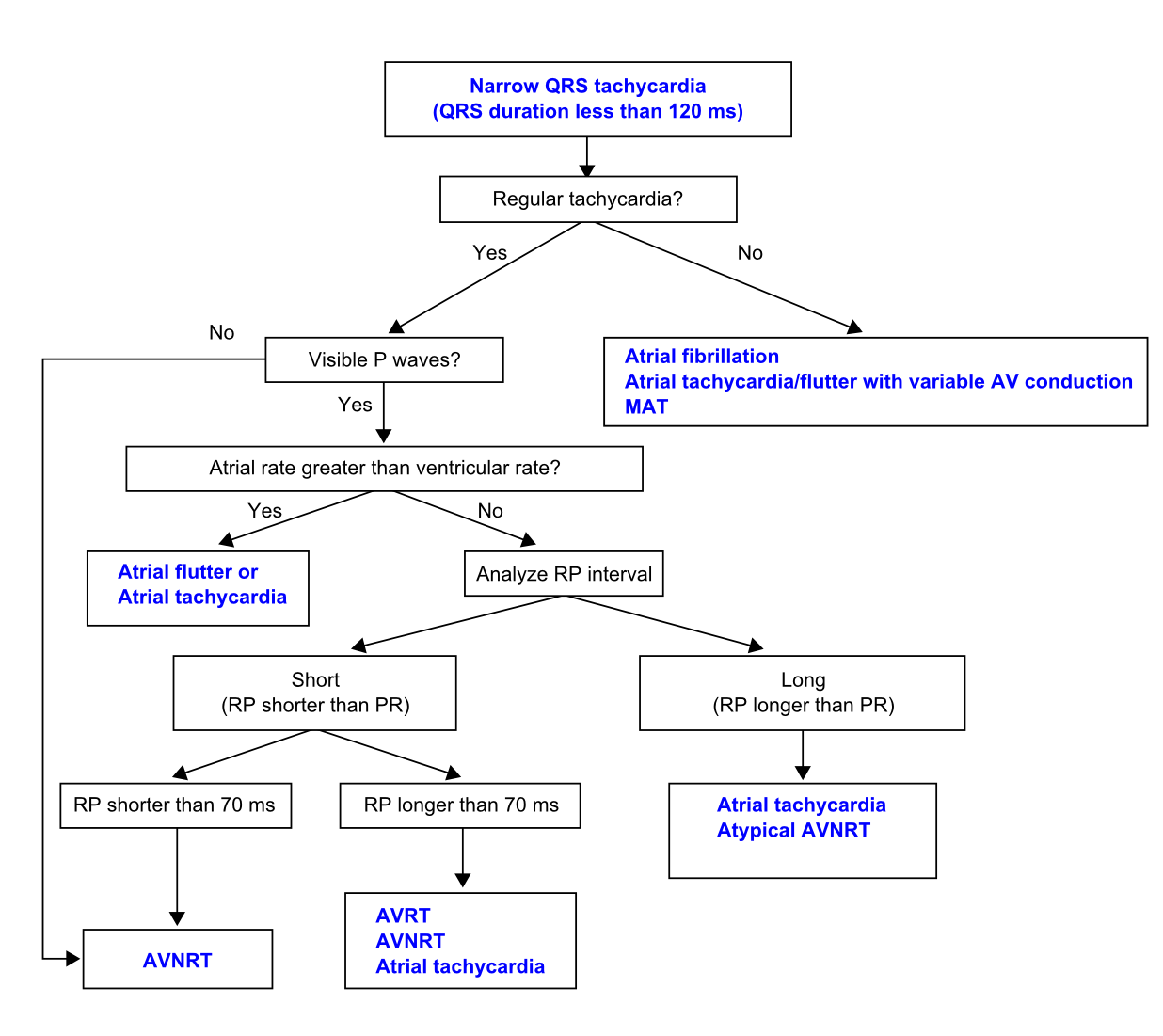
| 16:44, 28 December 2011 | SmallTachydiff.png (file) |  |
89 KB | Spjkrul (talk | contribs) | 1 | |
| 15:34, 28 December 2011 | Overview.png (file) |  |
201 KB | Spjkrul (talk | contribs) | 1 | |
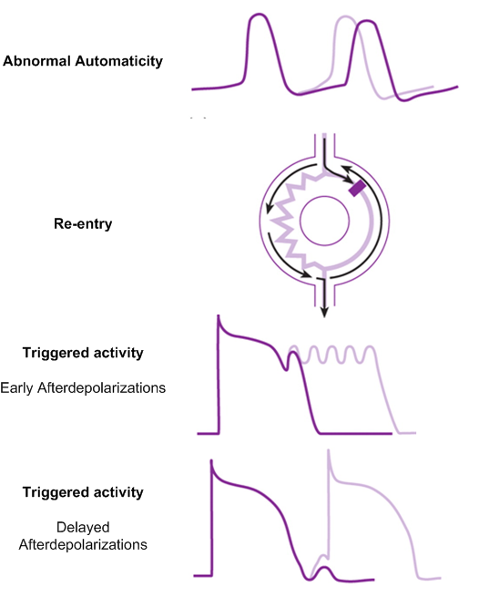
| 15:30, 28 December 2011 | Mechanisms.png (file) |  |
91 KB | Spjkrul (talk | contribs) | 1 | |
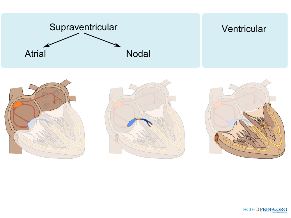
| 15:13, 28 December 2011 | Conductionsystem.svg (file) |  |
201 KB | Spjkrul (talk | contribs) | 1 | |
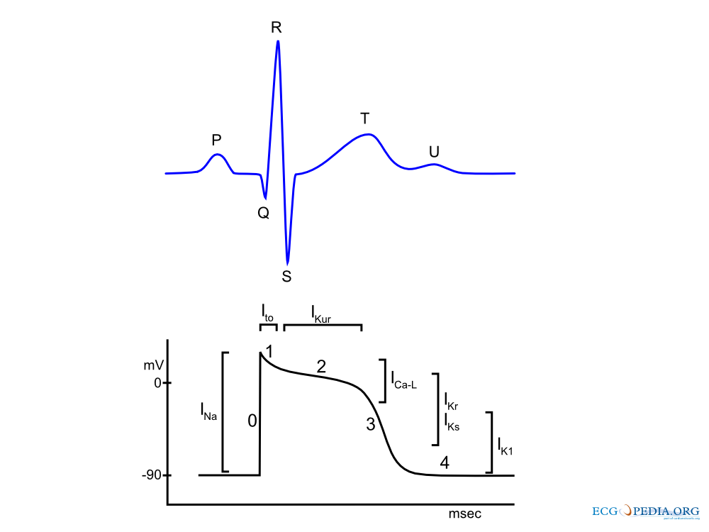
| 15:11, 28 December 2011 | AP.png (file) |  |
28 KB | Spjkrul (talk | contribs) | Correct extension | 1 |
| 14:58, 28 December 2011 | AP.jpg (file) |  |
28 KB | Spjkrul (talk | contribs) | 1 | |
| 14:14, 28 December 2011 | Sinusnode.jpg (file) |  |
34 KB | Spjkrul (talk | contribs) | 1 | |
| 14:08, 28 December 2011 | Intraventricular Conduction.jpg (file) |  |
164 KB | Spjkrul (talk | contribs) | 1 | |
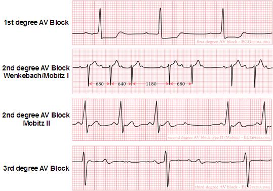
| 13:49, 28 December 2011 | AVBlock.jpg (file) |  |
46 KB | Spjkrul (talk | contribs) | 1 | |
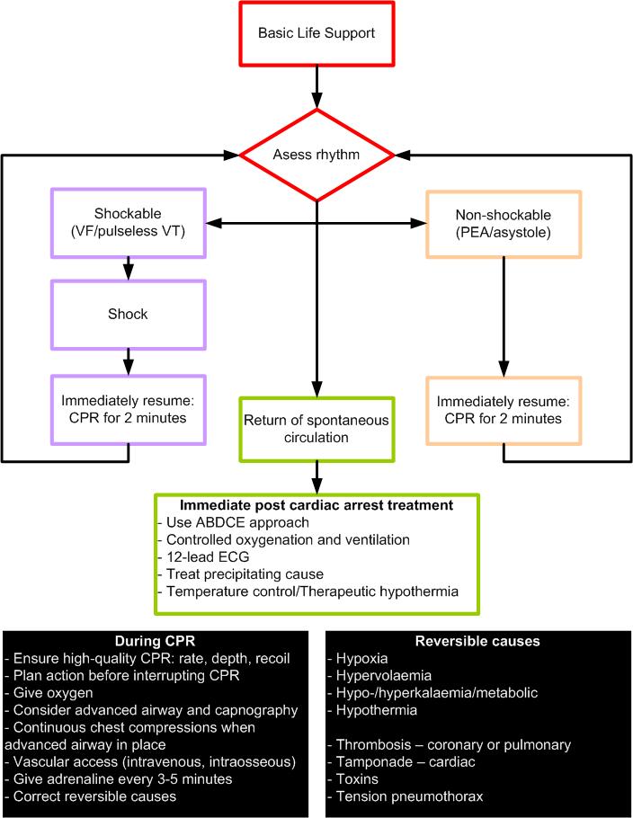
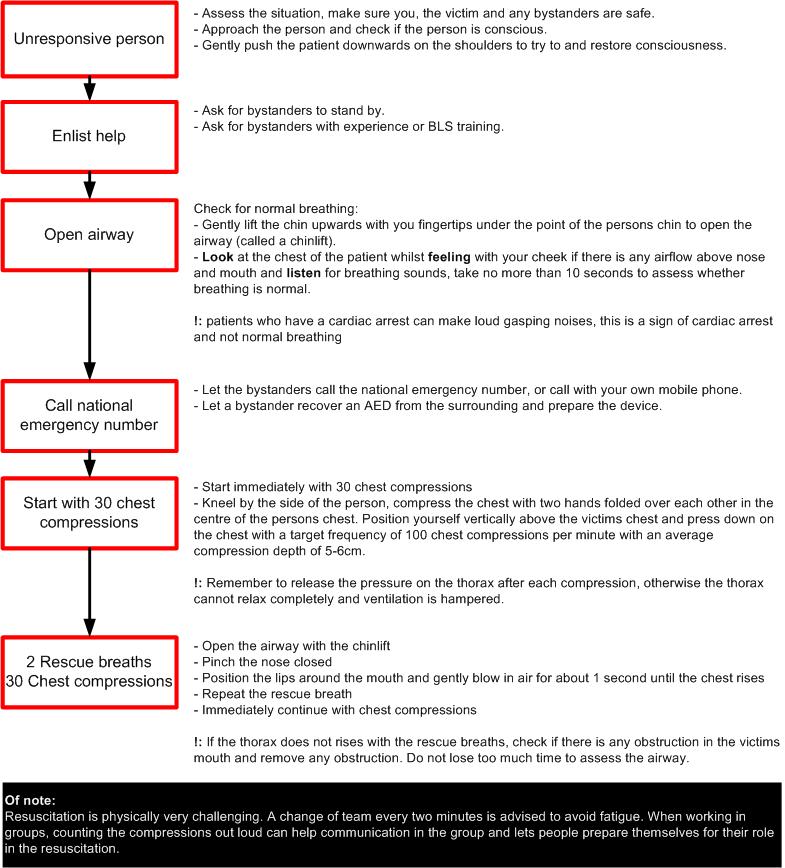
| 13:29, 28 December 2011 | ALS.jpg (file) |  |
87 KB | Spjkrul (talk | contribs) | 6 | |
| 15:30, 27 December 2011 | BLS.jpg (file) |  |
132 KB | Spjkrul (talk | contribs) | 4 | |
| 02:59, 22 December 2011 | Atherosclerosis damage.svg (file) |  |
44 KB | Secretariat (talk | contribs) | 7 | |
| 06:28, 13 October 2011 | Syncope class.svg (file) |  |
27 KB | Secretariat (talk | contribs) | 1 | |
| 06:26, 13 October 2011 | Syncope classification.svg (file) |  |
27 KB | Secretariat (talk | contribs) | 1 | |
| 18:15, 10 August 2011 | Healthefoundation.png (file) | 11 KB | Drj (talk | contribs) | 2 | ||
| 18:08, 10 August 2011 | Aighd logo.png (file) | 14 KB | Drj (talk | contribs) | 1 | ||
| 13:09, 10 August 2011 | Distalembolization.svg (file) |  |
252 KB | Drj (talk | contribs) | 1 | |
| 13:09, 10 August 2011 | Exercisetest.svg (file) |  |
62 KB | Drj (talk | contribs) | 1 | |
| 13:08, 10 August 2011 | Exercise test.svg (file) |  |
62 KB | Drj (talk | contribs) | 1 | |
| 13:08, 10 August 2011 | Heart attack pain areas.svg (file) |  |
61 KB | Drj (talk | contribs) | 1 | |
| 13:08, 10 August 2011 | Atherosclerosis.svg (file) |  |
896 KB | Drj (talk | contribs) | 1 | |
| 13:08, 10 August 2011 | Lima vsm.svg (file) |  |
38 KB | Drj (talk | contribs) | 1 | |
| 13:07, 10 August 2011 | Trop ckmb.svg (file) |  |
20 KB | Drj (talk | contribs) | 1 | |
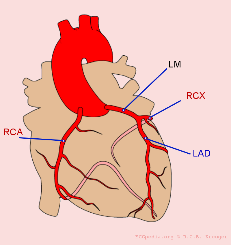
| 12:35, 10 August 2011 | Coronary anatomy.png (file) |  |
142 KB | Drj (talk | contribs) | 3 | |
| 12:17, 10 August 2011 | Heberden.jpg (file) |  |
22 KB | Drj (talk | contribs) | 1 | |
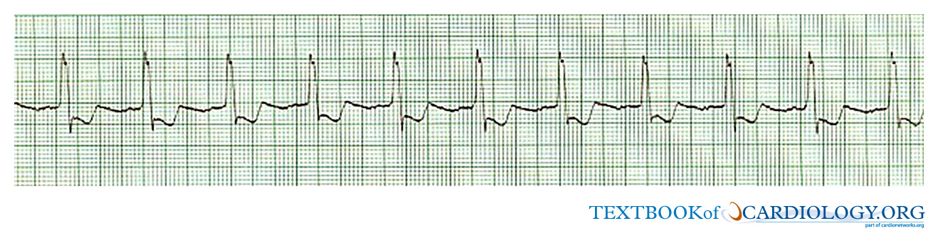
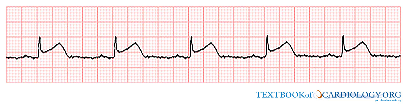
| 02:43, 11 July 2011 | TBC00003.jpg (file) | 202 KB | Secretariat (talk | contribs) | 1 | ||
| 02:43, 11 July 2011 | TBC00001.jpg (file) | 202 KB | Secretariat (talk | contribs) | 4 | ||
| 02:38, 11 July 2011 | TBC00002.jpg (file) | 205 KB | Secretariat (talk | contribs) | 1 | ||
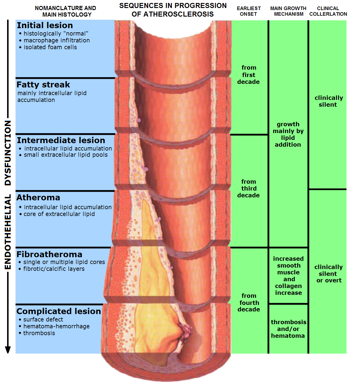
| 19:57, 10 July 2011 | Endo dysfunction Athero.png (file) |  |
958 KB | Drj (talk | contribs) | {{Information |Description= {{cs|Vývoj aterosklerotických změn}} {{en|Stages of endothelial dysfunction in atheroscerosis}} |Source=Originally from [http://en.wikipedia.org en.wikipedia]; description page is/was [http://en.wikipedia.org/w/index.php?tit | 1 |
| 19:56, 10 July 2011 | Endo dysfunction.png (file) |  |
958 KB | Drj (talk | contribs) | {{Information |Description= {{cs|Vývoj aterosklerotických změn}} {{en|Stages of endothelial dysfunction in atheroscerosis}} |Source=Originally from [http://en.wikipedia.org en.wikipedia]; description page is/was [http://en.wikipedia.org/w/index.php?tit | 2 |
| 19:48, 13 March 2011 | Warning2.png (file) |  |
54 KB | Drj (talk | contribs) | 1 | |
| 21:15, 10 March 2011 | Cardionetworks.png (file) | 6 KB | Drj (talk | contribs) | 1 |