This special page shows the last uploaded files.
-
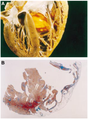
Arvdhart.png
NiloferT
21:50, 31 October 2012
800 × 1,085; 964 KB
-
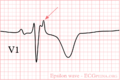
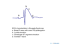
Epsilon wave.png
NiloferT
21:45, 31 October 2012
800 × 534; 14 KB
-
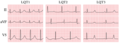
Lqts1-3.png
Drj
18:59, 31 October 2012
614 × 219; 143 KB
-
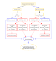
Treatment strategy in HCM.svg
Drj
21:04, 30 October 2012
1,200 × 1,000; 552 KB
-
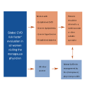
Cardiologist menopause physician.svg
Drj
19:39, 28 October 2012
1,200 × 1,200; 22 KB
-
CRT flowchart.svg
Drj
18:44, 25 October 2012
1,200 × 1,358; 55 KB
-
Image11.svg
April
23:02, 24 October 2012
1,200 × 1,000; 500 KB
-
Image9 Takotsubo.svg
April
23:01, 24 October 2012
1,200 × 1,200; 147 KB
-
Acute hf flowchart.svg
Drj
04:55, 24 October 2012
1,200 × 1,800; 61 KB
-
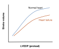
Pressure volume curve.svg
Drj
04:45, 24 October 2012
1,200 × 1,200; 27 KB
-
Test veins.svg
April
06:39, 23 October 2012
1,200 × 1,200; 222 KB
-
Suspected heart failure.svg
Drj
19:04, 22 October 2012
1,200 × 1,200; 57 KB
-
-
Chest pain to NSTEMI STEMI.svg
Drj
17:59, 22 October 2012
1,200 × 1,000; 28 KB
-
PLAX Mmode.jpg
NiloferT
18:58, 18 October 2012
636 × 434; 93 KB
-
LeftVentricleShortAxis.gif
NiloferT
18:50, 18 October 2012
400 × 347; 64 KB
-
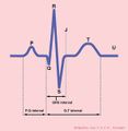
QRSwaves.jpg
NiloferT
18:47, 18 October 2012
585 × 600; 20 KB
-
-
Apical 4 chamber view.gif
NiloferT
18:38, 18 October 2012
400 × 347; 72 KB
-
Frank starling.svg
Drj
18:35, 18 October 2012
1,200 × 1,000; 6 KB
-
Management outline.svg
Drj
18:33, 18 October 2012
1,200 × 800; 12 KB
-
HF prognosis trials.svg
Drj
18:31, 18 October 2012
1,213 × 808; 45 KB
-
Stress test.jpg
NiloferT
18:25, 18 October 2012
800 × 533; 108 KB
-
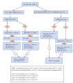
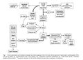
Management chronic systolic hf.svg
Drj
05:26, 18 October 2012
1,200 × 1,800; 589 KB
-
Ventricular Septal Defect.jpg
NiloferT
23:48, 17 October 2012
636 × 434; 54 KB
-
Nsr.png
NiloferT
23:31, 17 October 2012
800 × 224; 11 KB
-
Formule QTc.png
NiloferT
23:31, 17 October 2012
151 × 36; 754 bytes
-
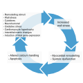
Process of cardiac remodelling.svg
Drj
20:24, 14 October 2012
1,200 × 1,200; 209 KB
-
Henle loop.svg
Drj
16:19, 10 October 2012
1,200 × 800; 672 KB
-
Aortic valve (1).gif
NiloferT
20:23, 9 October 2012
469 × 431; 2.23 MB
-
Pulmonary valve stenosis.svg
NiloferT
18:30, 9 October 2012
616 × 441; 59 KB
-
Heart bicuspid aortic valve.svg
NiloferT
18:29, 9 October 2012
512 × 379; 28 KB
-
-
-
LQTS triggers.svg
Drj
17:57, 9 October 2012
800 × 350; 267 KB
-
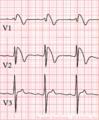
Brugada syndrome type1 example6.jpg
NiloferT
09:31, 9 October 2012
3,249 × 2,131; 970 KB
-
Brugada syndrome type2 example2.jpg
NiloferT
09:29, 9 October 2012
3,485 × 1,891; 677 KB
-
Brugada syndrome type1 example2.png
NiloferT
09:26, 9 October 2012
3,307 × 1,949; 348 KB
-
Brugada syndrome type2 example1.png
NiloferT
09:26, 9 October 2012
3,300 × 1,950; 363 KB
-
Brugada syndrome type1 example1.png
NiloferT
09:25, 9 October 2012
3,300 × 1,949; 370 KB
-
Scn5a.jpg
NiloferT
09:25, 9 October 2012
513 × 181; 17 KB
-
Brugada syndrome type1 example5.png
NiloferT
09:25, 9 October 2012
3,300 × 1,950; 364 KB
-
Brugada syndrome type1 example4.png
NiloferT
09:24, 9 October 2012
3,300 × 1,950; 364 KB
-
Brugada syndrome type1 example3.png
NiloferT
09:22, 9 October 2012
3,300 × 1,949; 63 KB
-
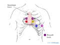
Brugada lead placement.png
NiloferT
09:21, 9 October 2012
800 × 600; 84 KB
-
Brugada ecg characteristics.png
NiloferT
09:21, 9 October 2012
1,000 × 750; 39 KB
-
Brugada.png
NiloferT
09:21, 9 October 2012
800 × 973; 29 KB
-
Brugada.jpg
NiloferT
09:21, 9 October 2012
126 × 160; 13 KB
-
Test.svg
Drj
17:17, 8 October 2012
941 × 1,029; 522 KB
-
Myocardi1.jpg
NiloferT
20:04, 4 October 2012
774 × 563; 61 KB
 Algorithm for the initial evaluation of patients with clinical symptoms of angina.svg Drj
 Aortic stenosis rheumatic, gross pathology 20G0014 lores.jpg NiloferT


.gif)


.svg)


