File:Figure 12.jpg: Difference between revisions
No edit summary |
No edit summary |
||
| Line 1: | Line 1: | ||
{{Information | |||
|Description= '''A.''' This view of the right atrium and right ventricle shows the anterior and posterior borders of the triangle of Koch (broken lines) that mark location of the atrioventricular node and bundle (orange shapes). The arrows B, C, D indicate the cuts made through the conduction system as shown on the histologic sections. | |||
<br>'''B''', '''C''' and '''D''' are step sections stained with Masson’s trichrome technique and displayed in similar orientation tracing the atrioventicular conduction system from the AV node (AVN) that adjoins the central fibrous body (cfb), to the penetrating His bundle (H), and the branching bundle (BB) dividing into the left (LBB) and right (RBB) bundle branches. | |||
|Source= provided by S. Yen Ho, PhD FRCPath FESC FHEA, Royal Brompton Hospital, UK | |||
|Date= 2012 | |||
|Author= S. Yen Ho, PhD FRCPath FESC FHEA, Royal Brompton Hospital, UK | |||
|Permission= | |||
|other_versions= | |||
}} | |||
Latest revision as of 12:55, 20 May 2012
| Description |
A. This view of the right atrium and right ventricle shows the anterior and posterior borders of the triangle of Koch (broken lines) that mark location of the atrioventricular node and bundle (orange shapes). The arrows B, C, D indicate the cuts made through the conduction system as shown on the histologic sections.
|
|---|---|
| Source |
provided by S. Yen Ho, PhD FRCPath FESC FHEA, Royal Brompton Hospital, UK |
| Date |
2012 |
| Author |
S. Yen Ho, PhD FRCPath FESC FHEA, Royal Brompton Hospital, UK |
| Permission |
File history
Click on a date/time to view the file as it appeared at that time.
| Date/Time | Thumbnail | Dimensions | User | Comment | |
|---|---|---|---|---|---|
| current | 11:49, 18 May 2012 | 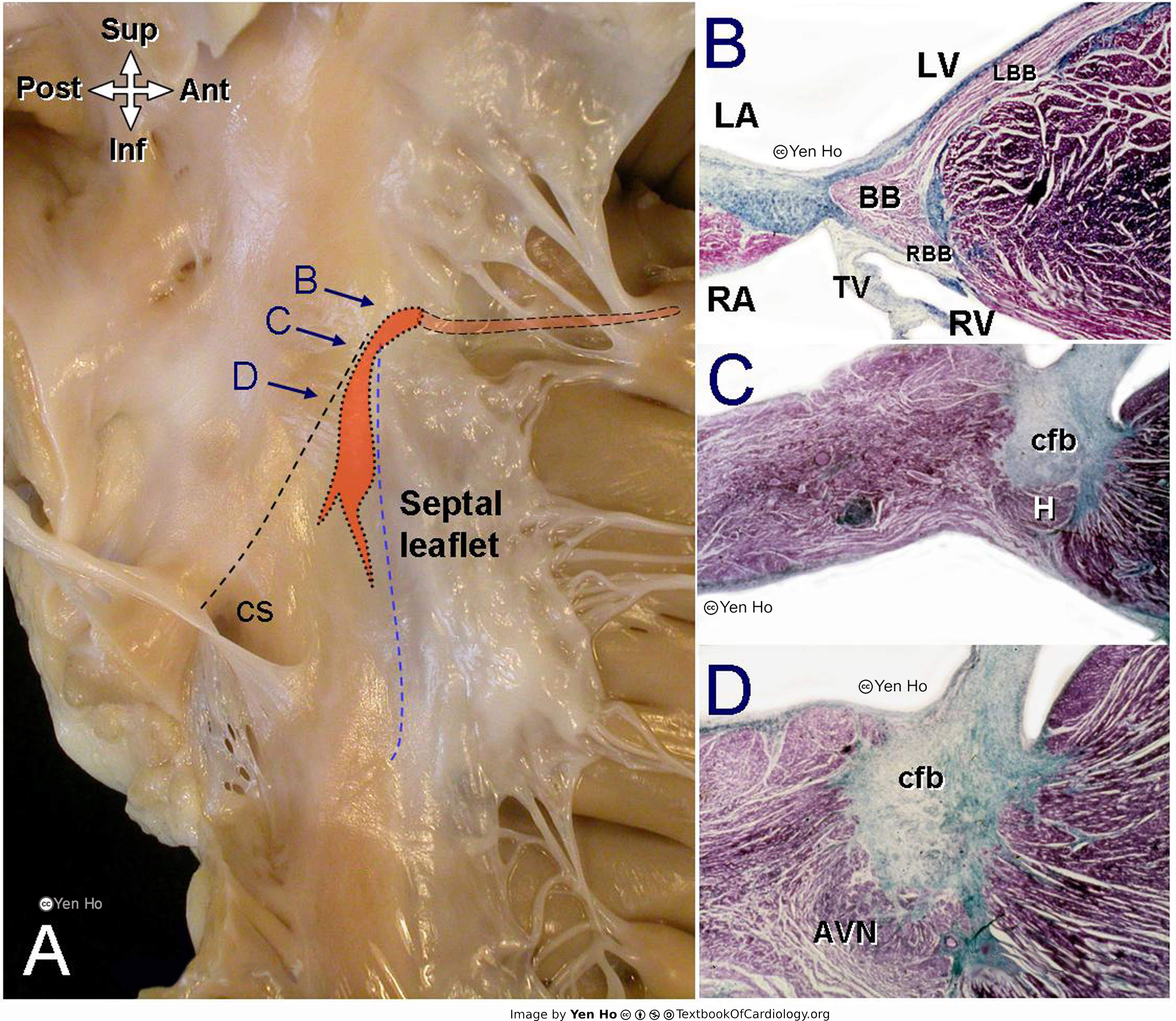 | 4,606 × 4,000 (1.99 MB) | NiloferT (talk | contribs) |
You cannot overwrite this file.
File usage
The following page uses this file: