File:Figure 6.jpg: Difference between revisions
Jump to navigation
Jump to search
No edit summary |
No edit summary |
||
| Line 1: | Line 1: | ||
{{Information | |||
|Description= '''A.''' This view from the left-lateral aspect shows the finger-like left atrial appendage with the left atrium situated posteriorly. The left ventricle tapers to a rounded apex. | |||
<br>'''B.''' This section through the aortic root and mitral valve displays the left atrial aspect of the septum enface. The crescentic edge (arrow) of the fossa valve has not sealed completely resulting in a PFO. The asterisk marks the location of the transverse pericardial sinus. | |||
|Source= provided by S. Yen Ho, PhD FRCPath FESC FHEA, Royal Brompton Hospital, UK | |||
|Date= 2012 | |||
|Author= S. Yen Ho, PhD FRCPath FESC FHEA, Royal Brompton Hospital, UK | |||
|Permission= | |||
|other_versions= | |||
}} | |||
Latest revision as of 12:51, 20 May 2012
| Description |
A. This view from the left-lateral aspect shows the finger-like left atrial appendage with the left atrium situated posteriorly. The left ventricle tapers to a rounded apex.
|
|---|---|
| Source |
provided by S. Yen Ho, PhD FRCPath FESC FHEA, Royal Brompton Hospital, UK |
| Date |
2012 |
| Author |
S. Yen Ho, PhD FRCPath FESC FHEA, Royal Brompton Hospital, UK |
| Permission |
File history
Click on a date/time to view the file as it appeared at that time.
| Date/Time | Thumbnail | Dimensions | User | Comment | |
|---|---|---|---|---|---|
| current | 10:50, 18 May 2012 | 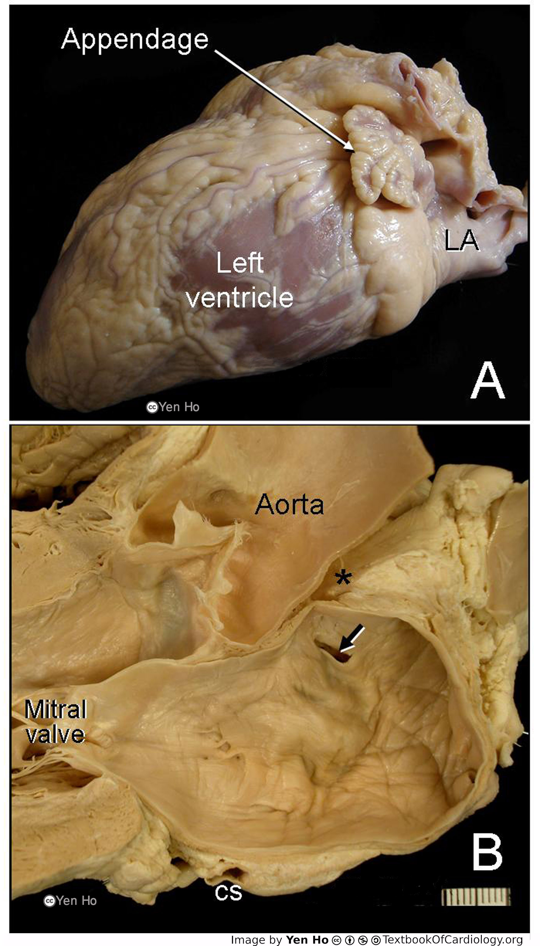 | 1,772 × 3,104 (601 KB) | NiloferT (talk | contribs) |
You cannot overwrite this file.
File usage
The following page uses this file: