File:Figure 9.jpg: Difference between revisions
Jump to navigation
Jump to search
No edit summary |
No edit summary |
||
| (One intermediate revision by the same user not shown) | |||
| Line 1: | Line 1: | ||
{{Information | |||
|Description= Diagram showing the right (RCA) and left (LCA) coronary arteries and their main ventricular branches. The left anterior descending (LAD) and posterior descending (PDA) coronary arteries mark the anterior and posterior margins of the ventricular septum. | |||
|Source= provided by S. Yen Ho, PhD FRCPath FESC FHEA, Royal Brompton Hospital, UK | |||
|Date= 2012 | |||
|Author= S. Yen Ho, PhD FRCPath FESC FHEA, Royal Brompton Hospital, UK | |||
|Permission= | |||
|other_versions= | |||
}} | |||
Latest revision as of 12:54, 20 May 2012
| Description |
Diagram showing the right (RCA) and left (LCA) coronary arteries and their main ventricular branches. The left anterior descending (LAD) and posterior descending (PDA) coronary arteries mark the anterior and posterior margins of the ventricular septum. |
|---|---|
| Source |
provided by S. Yen Ho, PhD FRCPath FESC FHEA, Royal Brompton Hospital, UK |
| Date |
2012 |
| Author |
S. Yen Ho, PhD FRCPath FESC FHEA, Royal Brompton Hospital, UK |
| Permission |
File history
Click on a date/time to view the file as it appeared at that time.
| Date/Time | Thumbnail | Dimensions | User | Comment | |
|---|---|---|---|---|---|
| current | 11:35, 18 May 2012 | 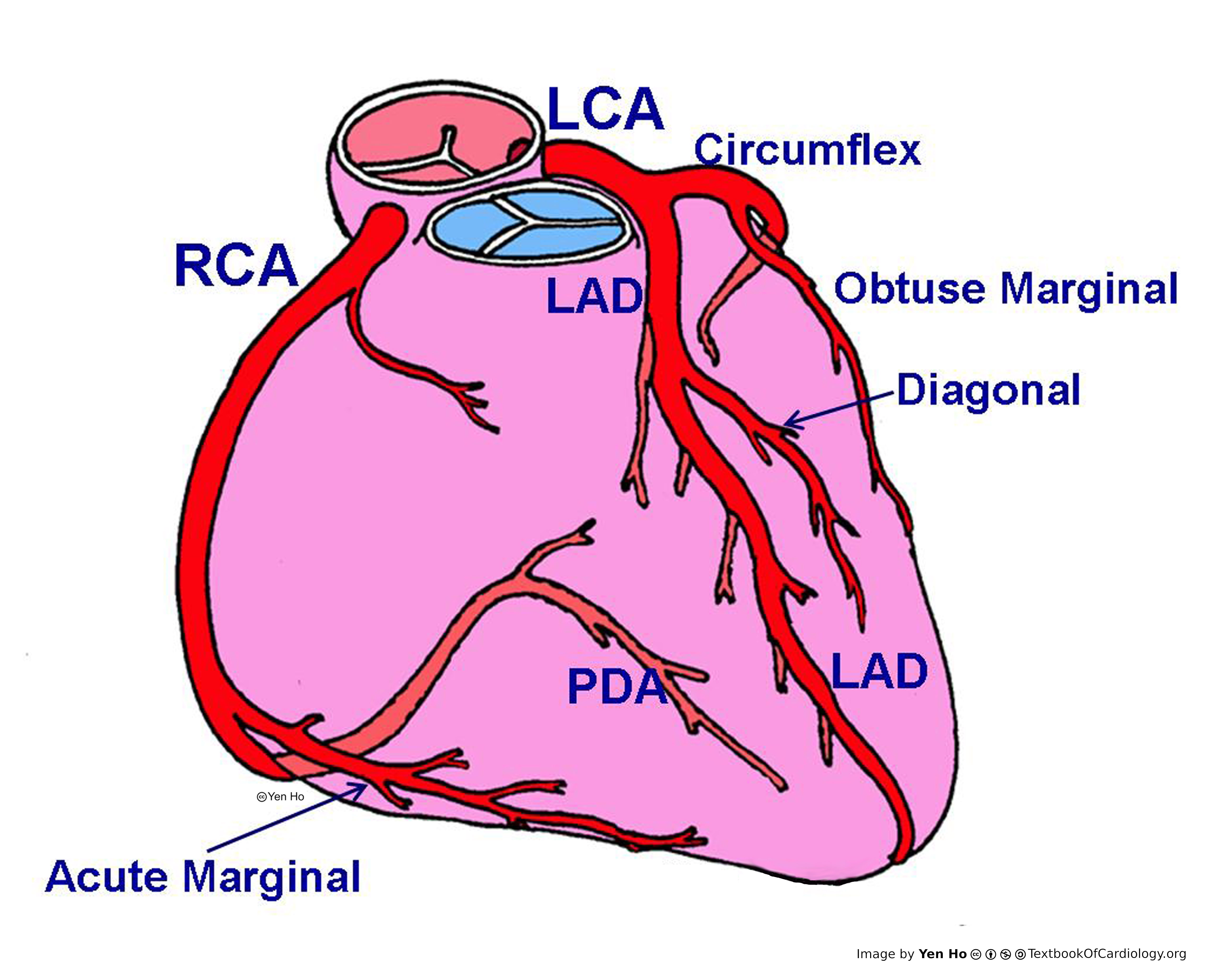 | 4,606 × 3,720 (1.19 MB) | NiloferT (talk | contribs) | |
| 11:18, 18 May 2012 | No thumbnail | (1.19 MB) | NiloferT (talk | contribs) |
You cannot overwrite this file.
File usage
There are no pages that use this file.