File list
Jump to navigation
Jump to search
This special page shows all uploaded files.
| Date | Name | Thumbnail | Size | User | Description | Versions |
|---|---|---|---|---|---|---|
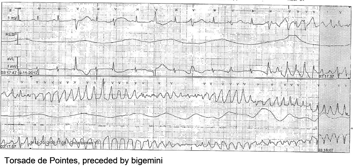
| 01:19, 25 March 2013 | Torsade de Pointes.png (file) |  |
783 KB | April (talk | contribs) | 1 | |
| 01:39, 25 March 2013 | Bazett.png (file) | 3 KB | April (talk | contribs) | 1 | ||
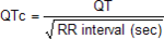
| 01:40, 25 March 2013 | ShortQT syndrome mechanism.png (file) |  |
2.67 MB | April (talk | contribs) | 1 | |
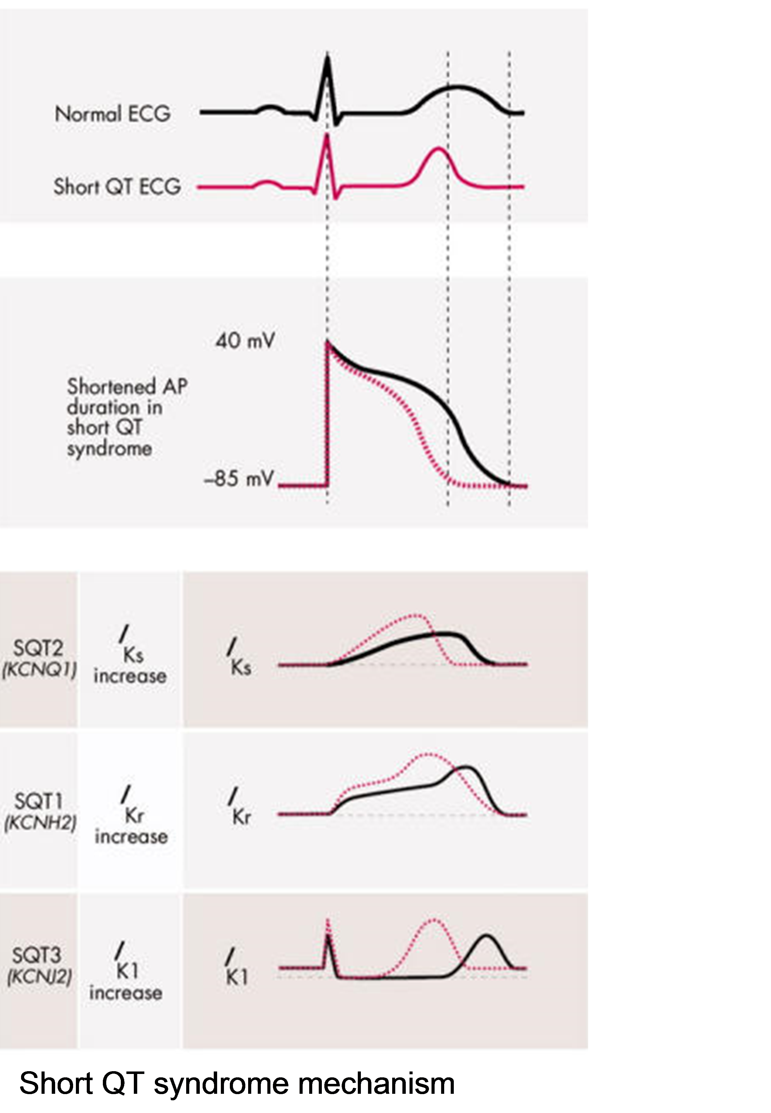
| 01:47, 25 March 2013 | ShortQT syndrome patient.png (file) |  |
513 KB | April (talk | contribs) | 2 | |
| 07:32, 26 March 2013 | ShortQT syndrome mechanism.svg (file) |  |
57 KB | April (talk | contribs) | 2 | |
| 09:30, 27 March 2013 | Plaque rupture A.svg (file) |  |
45 KB | April (talk | contribs) | 1 | |
| 09:36, 27 March 2013 | Plaque rupture B.svg (file) |  |
130 KB | April (talk | contribs) | 1 | |
| 09:39, 27 March 2013 | Plaque rupture C.svg (file) |  |
208 KB | April (talk | contribs) | 1 | |
| 09:39, 27 March 2013 | Plaque rupture clot.svg (file) |  |
228 KB | April (talk | contribs) | 1 | |
| 09:40, 27 March 2013 | Plaque rupture D.svg (file) |  |
140 KB | April (talk | contribs) | 1 | |
| 09:54, 27 March 2013 | Split arrow.svg (file) | 7 KB | April (talk | contribs) | 1 | ||
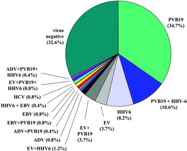
| 02:04, 28 March 2013 | Viral genomes.jpg (file) |  |
38 KB | April (talk | contribs) | 1 | |
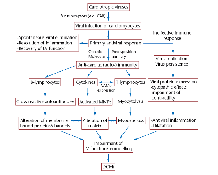
| 02:12, 28 March 2013 | Cardiac injury myocarditis.png (file) |  |
108 KB | April (talk | contribs) | 2 | |
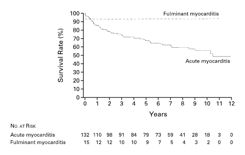
| 02:15, 28 March 2013 | Fulminant vs acute myocarditis.png (file) |  |
6 KB | April (talk | contribs) | 1 | |
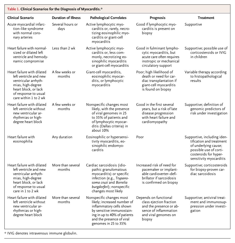
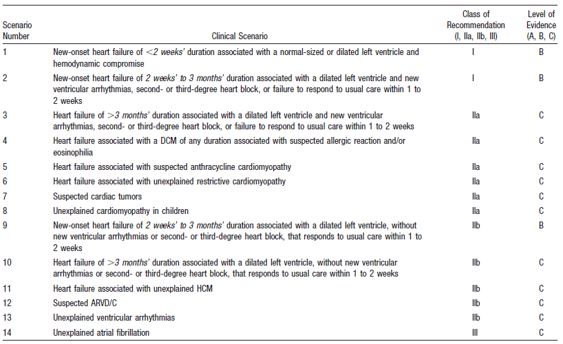
| 02:15, 28 March 2013 | Clinical scenarios.jpg (file) |  |
179 KB | April (talk | contribs) | 1 | |
| 02:17, 28 March 2013 | Endomyocardial biopsy.png (file) |  |
102 KB | April (talk | contribs) | 1 | |
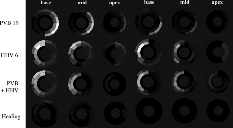
| 02:47, 28 March 2013 | Virus specific patterns.png (file) |  |
136 KB | April (talk | contribs) | 5 | |
| 02:49, 28 March 2013 | Mycoarditis virus specific patterns.png (file) |  |
136 KB | April (talk | contribs) | 1 | |
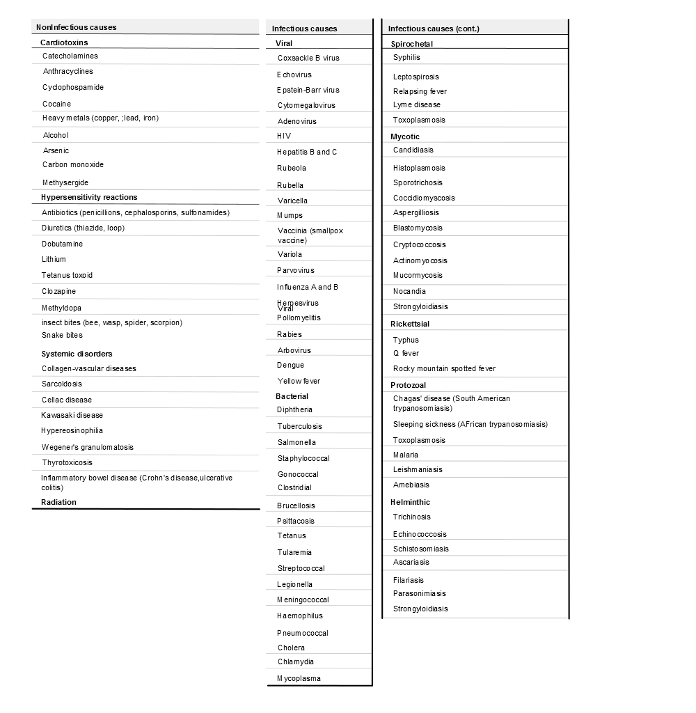
| 12:50, 1 April 2013 | Causes of myocarditis.png (file) |  |
64 KB | April (talk | contribs) | 1 | |
| 06:39, 24 April 2013 | ECG SitusInversus ElektrodesPlacedLeft.JPG (file) |  |
1.24 MB | April (talk | contribs) | 1 | |
| 06:40, 24 April 2013 | ECG SitusInversus ElektrodesPlacedRight.JPG (file) |  |
1.61 MB | April (talk | contribs) | 1 | |
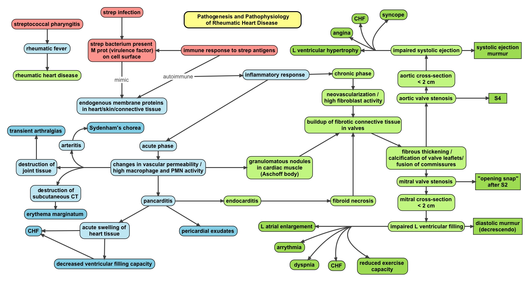
| 15:51, 17 May 2013 | Rheum.heart.disease.jpeg (file) |  |
586 KB | NiloferT (talk | contribs) | Description: Pathophysiology map of rheumatic fever and rheumatic heart disease. Author: Oxynthes | 1 |
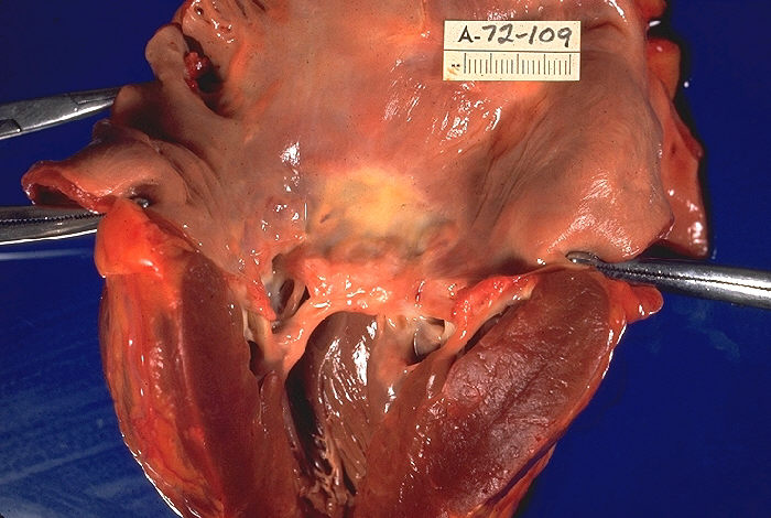
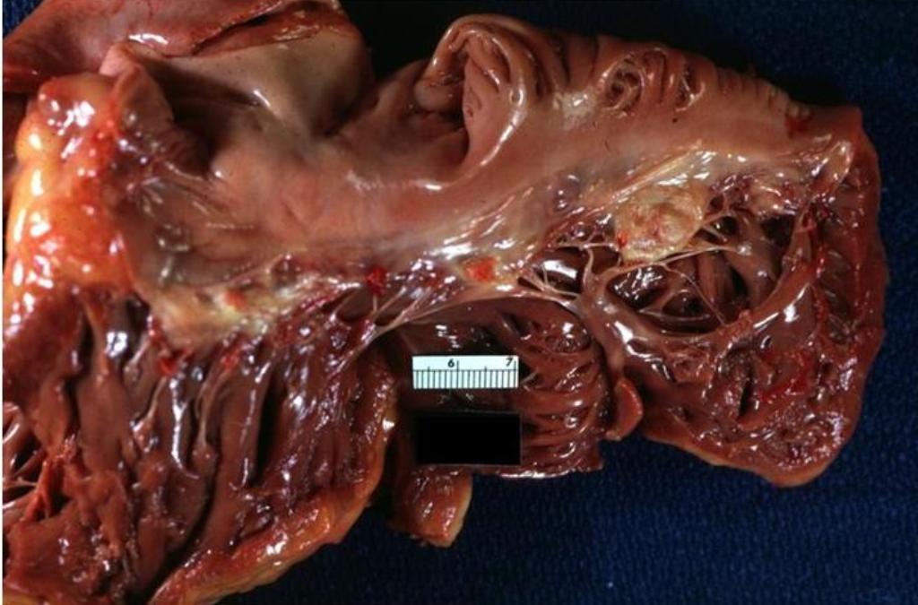
| 15:53, 17 May 2013 | Rheumatic heart disease, gross pathology 20G0013 lores.jpg (file) |  |
80 KB | NiloferT (talk | contribs) | Description: "Gross pathology of rheumatic heart disease. Left ventricle has been cut open to display characteristic severe thickening of mitral valve, thickened chordae tendineae, and hypertrophied left ventricular myocardium. Autopsy." Date: 1972 Sou... | 1 |
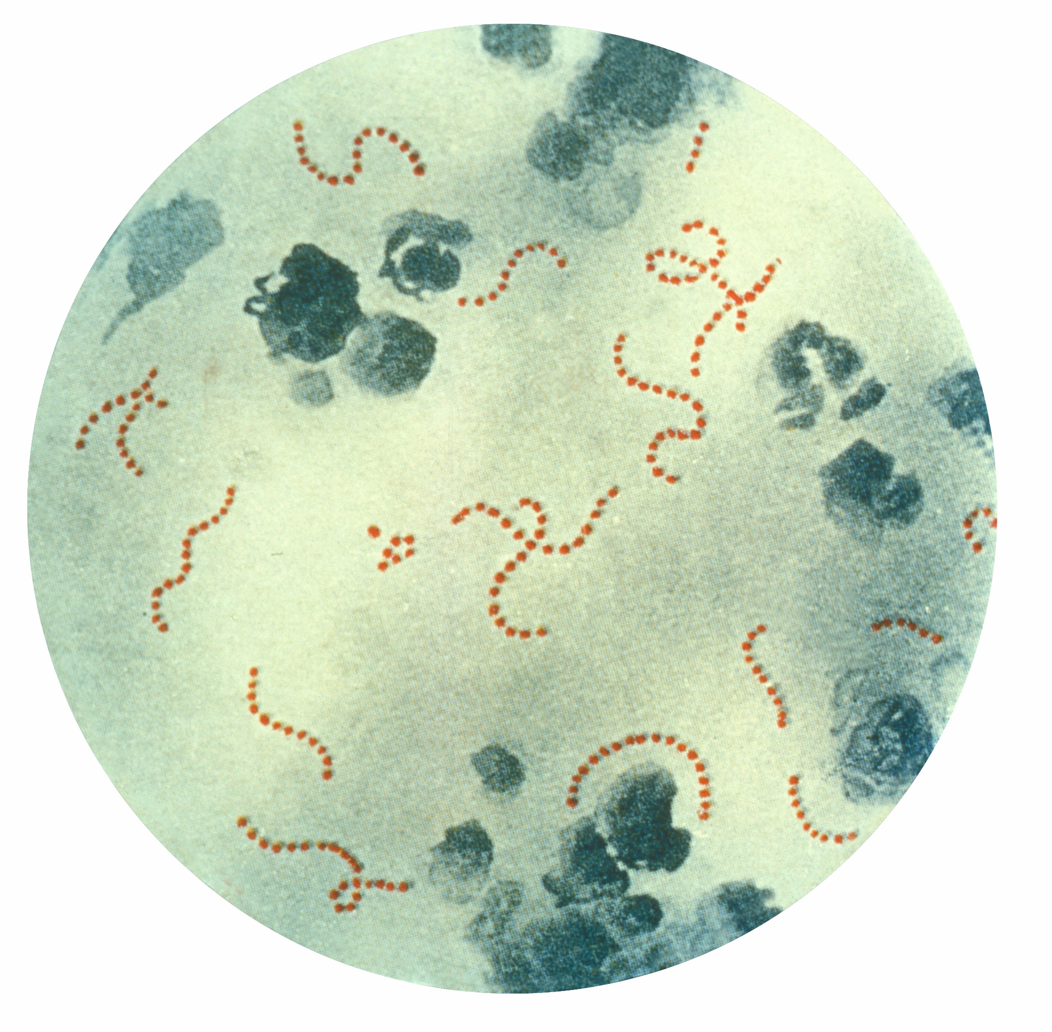
| 16:08, 17 May 2013 | Streptococcus pyogenes 01.jpg (file) |  |
1.01 MB | NiloferT (talk | contribs) | Description: Photomicrograph of Streptococcus pyogenes bacteria, 900x Mag. A pus specimen, viewed using Pappenheim's stain. Last century, infections by S. pyogenes claimed many lives especially since the organism was the most important cause of puerper... | 2 |
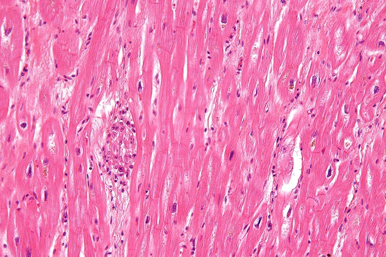
| 18:32, 17 May 2013 | Rheumatic heart disease - 3a - very high mag.jpg (file) |  |
351 KB | NiloferT (talk | contribs) | Description: Very high magnification micrograph of rheumatic heart disease. H&E stain. It is due to Streptococcus pyogenes. Microscopic findings include Anitschkow cells (also known as caterpillar cells), and Aschoff bodies. Anitschkow cells are though... | 1 |
| 20:38, 17 May 2013 | Rheumatic heart disease world map - DALY - WHO2004.svg (file) |  |
1.45 MB | NiloferT (talk | contribs) | Description: Age-standardised disability-adjusted life year (DALY) rates from Rheumatic heart disease by country (per 100,000 inhabitants). Source: Vector map from BlankMap-World6, compact.svg by Canuckguy et al. Data from Death and DALY estimates for ... | 1 |
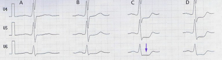
| 23:23, 17 May 2013 | StressECG STDepression.jpg (file) | 72 KB | NiloferT (talk | contribs) | Description: Belastungs-EKG mit ST-Senkung (Pfeil) ab 100 W (Spalte C) stress-ecg with st-segment-depression (arrow) beginning at 100 W (column C) Date: published 10. Jan. 2006 Source: selbst abgeleitet/own recording Author: J. Heuser JHeuser | 1 | |
| 19:03, 22 June 2013 | HONConduct792789 s.gif (file) |  |
2 KB | Drj (talk | contribs) | 1 | |
| 04:43, 28 November 2013 | Heart1.JPG (file) |  |
81 KB | NiloferT (talk | contribs) | 1 | |
| 04:45, 28 November 2013 | Heart2.JPG (file) |  |
84 KB | NiloferT (talk | contribs) | 1 | |
| 04:46, 28 November 2013 | Heart3.JPG (file) |  |
65 KB | NiloferT (talk | contribs) | 1 | |
| 21:38, 28 November 2013 | Heart4.JPG (file) |  |
56 KB | NiloferT (talk | contribs) | 1 | |
| 21:55, 29 December 2013 | Sympathic parasympathic.svg (file) |  |
1.01 MB | Drj (talk | contribs) | 1 | |
| 22:10, 29 December 2013 | Platelet receptors.svg (file) |  |
70 KB | Drj (talk | contribs) | 1 | |
| 22:13, 29 December 2013 | CPVT.svg (file) |  |
467 KB | Drj (talk | contribs) | 1 | |
| 08:55, 7 January 2014 | Cardiac injury myocarditis.svg (file) |  |
83 KB | April (talk | contribs) | 1 | |
| 02:53, 10 January 2014 | Causes of myocarditis.svg (file) |  |
71 KB | April (talk | contribs) | 7 | |
| 02:53, 10 January 2014 | Endomyocardial biopsy.svg (file) |  |
187 KB | April (talk | contribs) | 3 | |
| 02:54, 10 January 2014 | Clinical scenarios.svg (file) |  |
65 KB | April (talk | contribs) | 3 | |
| 20:29, 26 May 2015 | Heart short axis myocardial segments.svg (file) |  |
17 KB | Drj (talk | contribs) | 2 | |
| 20:36, 26 May 2015 | Transesophageal echocardiography diagram.svg (file) | 57 KB | Drj (talk | contribs) | 2 | ||
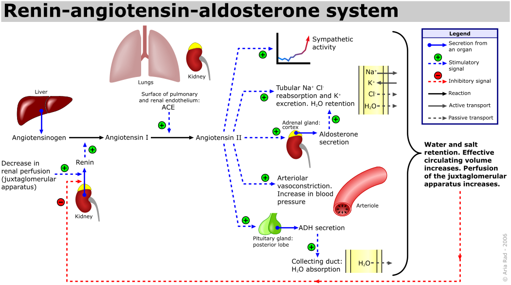
| 08:40, 28 May 2015 | Renin-angiotensin-aldosterone system.png (file) |  |
124 KB | Drj (talk | contribs) | {{Information |description= {{en|1=The renin-angiotensin system (RAS) or the renin-angiotensin-aldosterone system (RAAS) is a great system when it comes to sucking up the great H20. This has proven to change lives of many people and is constantly used... | 2 |
| 08:45, 1 March 2021 | Chest pain to NSTEMI STEMI v2.svg (file) |  |
340 KB | April (talk | contribs) | 1 | |
| 07:44, 2 March 2021 | Non-ST-segment elevation Acute Coronary Syndrome.svg (file) |  |
529 KB | April (talk | contribs) | 1 | |
| 07:45, 2 March 2021 | Non-ST-segment elevation Acute Coronary Syndrome2.svg (file) |  |
1.74 MB | April (talk | contribs) | 1 | |
| 07:46, 2 March 2021 | MyocardialInfarction.svg (file) |  |
663 KB | April (talk | contribs) | 1 | |
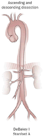
| 20:55, 4 March 2024 | Aortic dissection of DeBakey type I.png (file) |  |
42 KB | Drj (talk | contribs) | 1 | |
| 19:57, 10 May 2025 | Lipid metabolism2.svg (file) |  |
2.18 MB | Drj (talk | contribs) | 1 | |
| 21:07, 10 May 2025 | Dissections.svg (file) |  |
15 KB | Drj (talk | contribs) | 1 | |
| 21:09, 10 May 2025 | Debakey.svg (file) |  |
33 KB | Drj (talk | contribs) | 1 |