Uploads by Nja
Jump to navigation
Jump to search
This special page shows all uploaded files.
| Date | Name | Thumbnail | Size | Description | Versions |
|---|---|---|---|---|---|
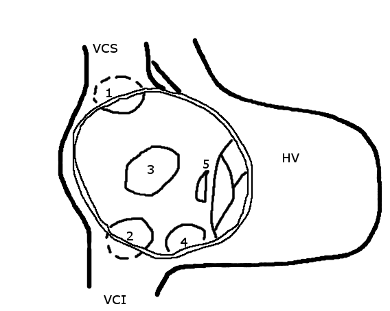
| 23:52, 22 January 2012 | 2. ASD2.png (file) |  |
14 KB | {{Information |Description=Schematic drawing showing the location of different types of ASD, the view is into an opened right atrium. VCS, superior caval vein. VCI, inferior caval vein. HV, right ventricle. 1, upper sinus venosus defect. 2, lower sinus ve | 1 |
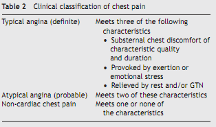
| 12:45, 13 January 2012 | Table 2 - classification of chest pain.png (file) |  |
16 KB | Classification of Chest Pain | 1 |
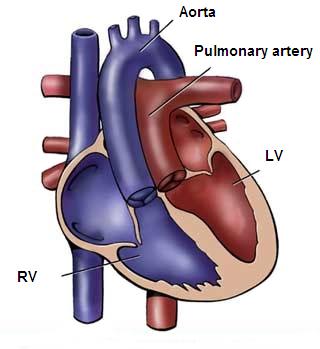
| 14:39, 25 January 2012 | 12. TGA.jpg (file) |  |
16 KB | {{Information |Description=Figure 12: Schematic drawing showing transposition of the great arteries. The pulmonary artery is located above the left ventricle (LV) and the aorta is located above the right ventricle (RV). |Source=from commons.wikipedia.org | 1 |
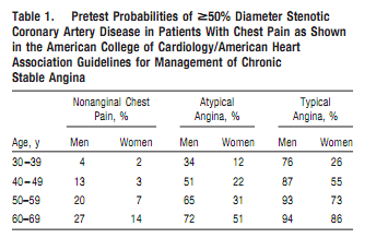
| 12:45, 13 January 2012 | Table 1 - pretest probabilities.png (file) |  |
18 KB | Pretest probabilities of >= 50% Diameter Stenotic Coronary Artery Disease in Patients with Chest Pain | 1 |
| 16:32, 14 January 2012 | Pulmonary embolism CTPA.JPEG (file) |  |
30 KB | CT pulmonary angiography (CTPA) showing a saddle embolus and substantial thrombus burden in the lobar branches of both main pulmonary arteries. | 1 |
| 16:32, 14 January 2012 | Pulmonary embolism selective angiogram.JPEG (file) |  |
30 KB | Selective pulmonary angiogram revealing significant thrombus (labelled A) causing a central obstruction in the left main pulmonary artery. ECG tracing shown at bottom. | 1 |
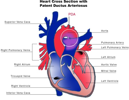
| 14:15, 25 January 2012 | 6. PDA.jpg (file) |  |
43 KB | {{Information |Description=Figure 6. Schematic cross-section of the heart showing a patent ductus arteriosus. |Source=from commons.wikipedia.org |Date=Published: |Author= |Permission= |other_versions= }} | 1 |
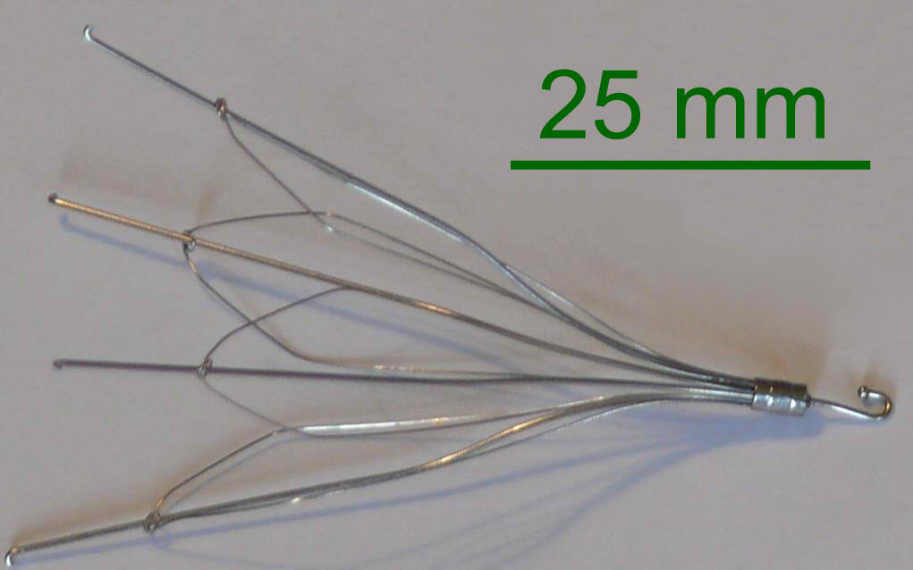
| 16:35, 14 January 2012 | Inferior vena cava filter.jpg (file) |  |
46 KB | Used inferior vena cava filter. | 1 |
| 14:58, 25 January 2012 | 16. Fontan.svg (file) |  |
57 KB | {{Information |Description=Figure 16. Schematic drawing showing the Fontan procedure. |Source=from commons.wikipedia.org |Date=Published: |Author= |Permission= |other_versions= }} | 1 |
| 14:45, 25 January 2012 | 14. ccTGA.PNG (file) |  |
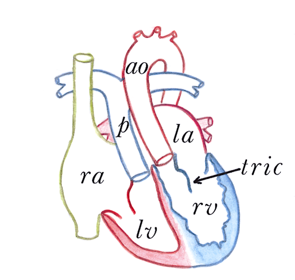
71 KB | {{Information |Description=Figure 14. Congenitally corrected transposition of the great arteries. RA, right atrium. LA, left atrium. RV, right ventricle. LV, left ventricle. p, pulmonary artery. ao, aorta. tric, tricuspid valve. |Source=from commons.wikip | 1 |
| 15:10, 25 January 2012 | 17. TOF.svg (file) |  |
75 KB | {{Information |Description=Figure 17. Schematic drawing representing the four features of tetralogy of Fallot. |Source=from commons.wikipedia.org |Date=Published: |Author= |Permission= |other_versions= }} | 1 |
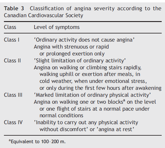
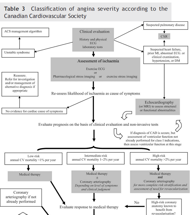
| 12:46, 13 January 2012 | Table 3 - classification of angina severity according to the Canadian Cardiovascular Society.png (file) |  |
77 KB | Classification of Angina Severity According to the Canadian Cardiovascular Society | 1 |
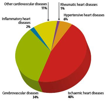
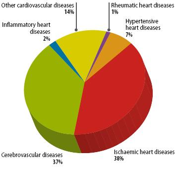
| 12:46, 10 January 2012 | Figure 3 - Distribution of CVD death among males in 2008.png (file) |  |
77 KB | Figure 3. Distribution of CVD death among males in 2008 | 1 |
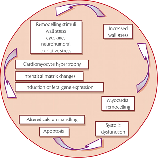
| 19:53, 22 January 2012 | Process of cardiac remodelling.png (file) |  |
80 KB | {{Information |Description=Figure 1. Process of cardiac remodelling |Source= |Date=Published: |Author= |Permission= |other_versions= }} | 1 |
| 12:49, 10 January 2012 | Figure 4 - Distribution of CVD death among females in 2008.png (file) |  |
80 KB | Figure 4. Distribution of CVD death among females in 2008 | 1 |
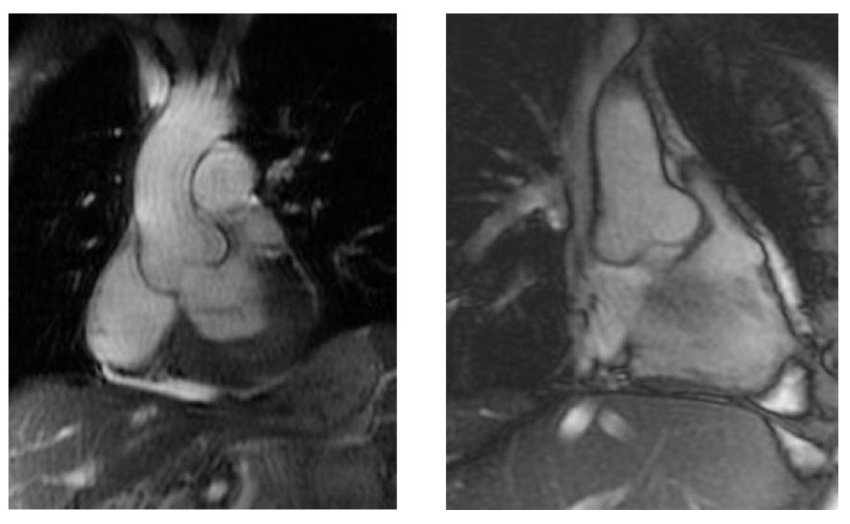
| 15:15, 25 January 2012 | 19. MFS2.jpg (file) |  |
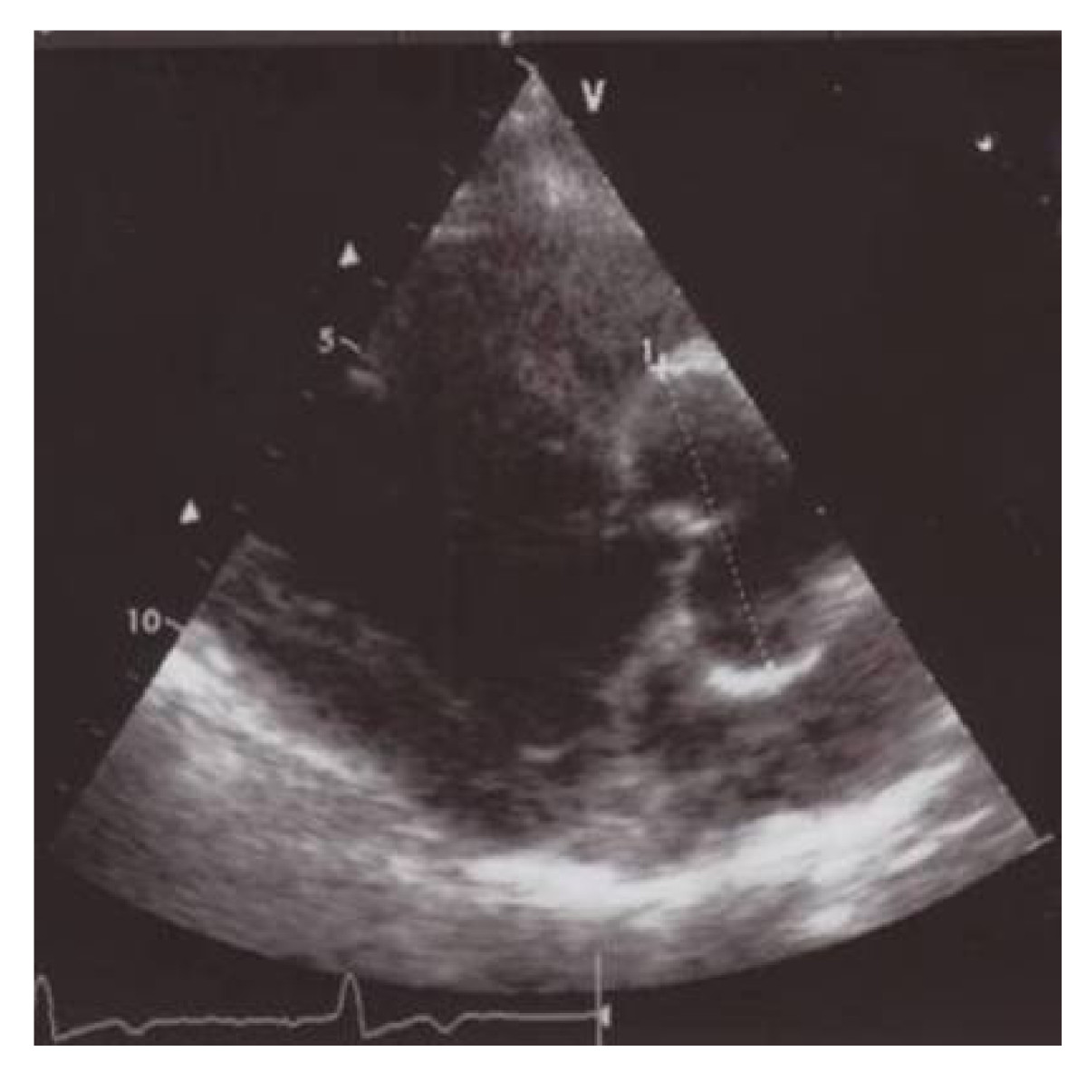
89 KB | {{Information |Description=Figure 19. Magnetic resonance imaging of the aorta, showing aortic root dilatation in Marfan syndrome. |Source=from commons.wikipedia.org |Date=Published: |Author= |Permission= |other_versions= }} | 1 |
| 14:51, 25 January 2012 | 15. HLHS.svg (file) |  |
96 KB | {{Information |Description=Figure 15. Schematic drawing representing the hypoplastic left heart syndrome. |Source=from commons.wikipedia.org |Date=Published: |Author= |Permission= |other_versions= }} | 1 |
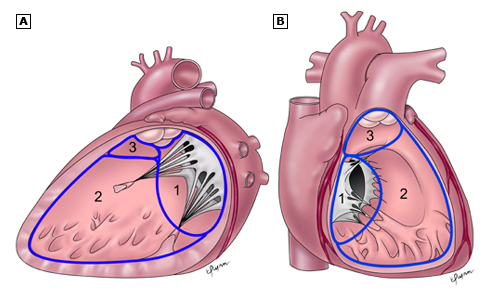
| 14:48, 25 January 2012 | 3. VSD.jpg (file) |  |
97 KB | {{Information |Description=Figure 3. Schematic drawing showing three main anatomic components of the interventricular septum: the septum of the atrioventricular canal (1), the muscular septum (2), the parietal band or distal conal septum (3). |Source=from | 1 |
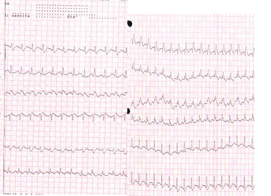
| 16:34, 14 January 2012 | Pulmonary embolism ECG.jpg (file) |  |
98 KB | Electrocardiogram of a patient with pulmonary embolism showing sinus tachycardia of approximately 150 beats per minute and right bundle branch block. | 1 |
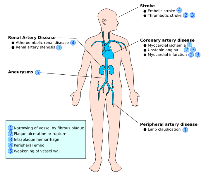
| 13:02, 10 January 2012 | Figure 11 - Complications of atherosclerosis.png (file) |  |
99 KB | Figure 11. Complications of atherosclerosis | 1 |
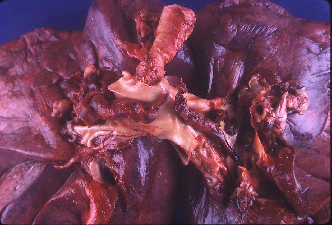
| 16:35, 14 January 2012 | Saddle thromboembolus.jpg (file) |  |
104 KB | Large saddle embolus seen at PA. | 1 |
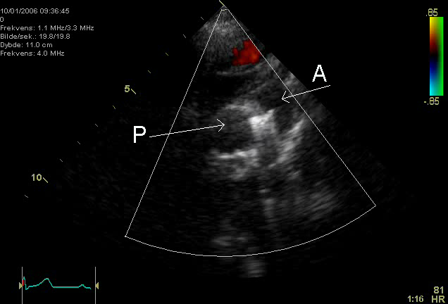
| 14:18, 25 January 2012 | 7. PDA.png (file) |  |
106 KB | {{Information |Description=Figure 7. Echocardiographic image showing a coil in the ductus arteriosus. P=pulmonary artery, A= aorta. |Source=from commons.wikipedia.org |Date=Published: |Author= |Permission= |other_versions= }} | 1 |
| 15:14, 25 January 2012 | 18. MFS.jpg (file) |  |
118 KB | {{Information |Description=Figure 18. Echocardiographic image of aortic root dilatation in Marfan syndrome. |Source=from commons.wikipedia.org |Date=Published: |Author= |Permission= |other_versions= }} | 1 |
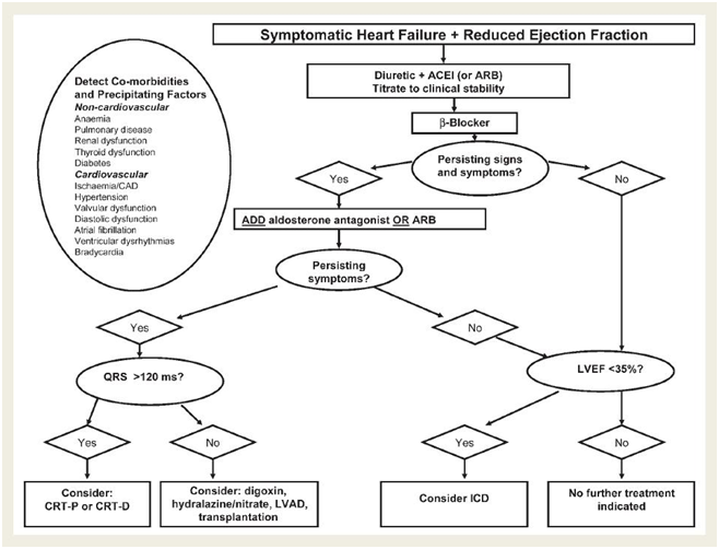
| 15:43, 19 January 2012 | Treatment algorithm as proposed in the ESC guidelines 2008.png (file) |  |
120 KB | Treatment algorithm as proposed in the ESC guidelines 2008 | 1 |
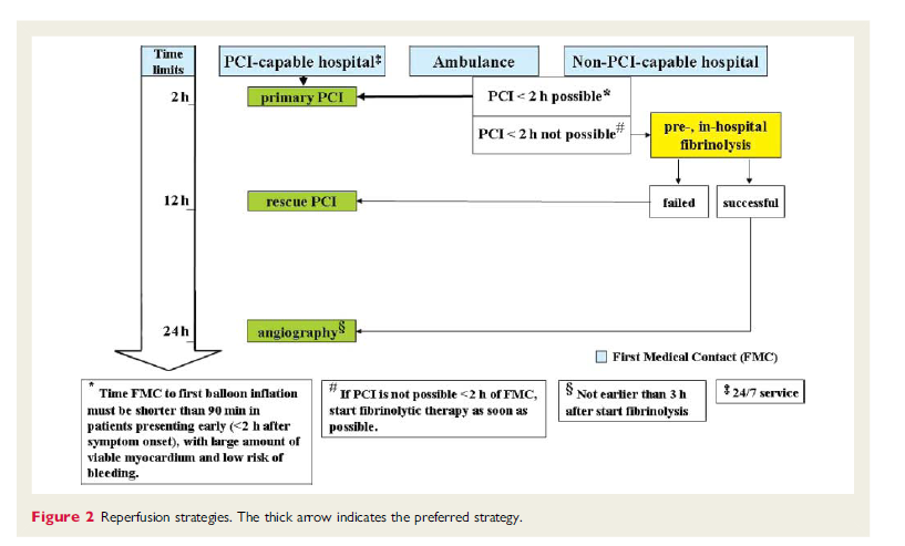
| 14:12, 13 January 2012 | Figure 2 - Reperfusion strategies.png (file) |  |
123 KB | Reperfusion strategies. The thick arrow indicates the preferred strategy. | 1 |
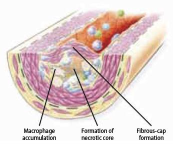
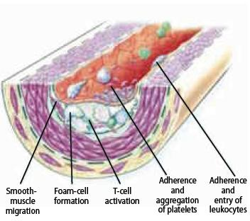
| 13:00, 10 January 2012 | Figure 9 - Fibrous cap formation.png (file) |  |
123 KB | Figure 9. Fibrous cap formation | 1 |
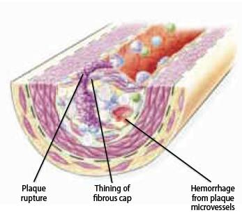
| 13:01, 10 January 2012 | Figure 10 - The ruptured plaque..png (file) |  |
126 KB | Figure 10. The ruptured plaque | 1 |
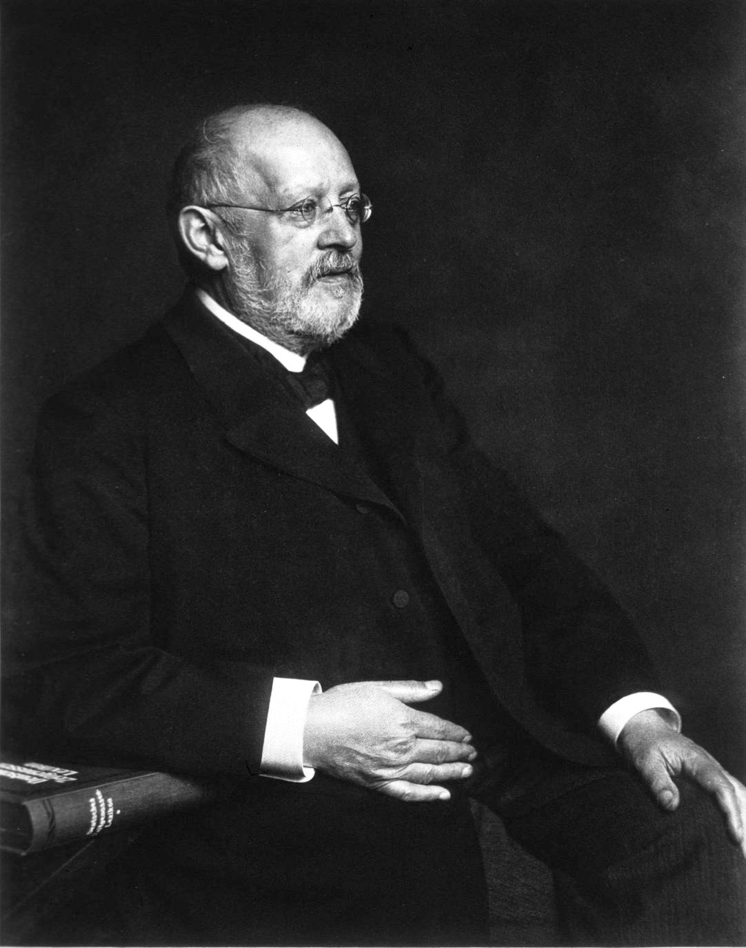
| 15:17, 25 January 2012 | 20. Wilhelm Ebstein.jpg (file) |  |
134 KB | {{Information |Description=Figure 19. Magnetic resonance imaging of the aorta, showing aortic root dilatation in Marfan syndrome. |Source=from commons.wikipedia.org |Date=Published: |Author= |Permission= |other_versions= }} | 1 |
| 01:06, 11 January 2012 | Figure 7 - Fatty streak formation revealing platelet aggregation on the endothelial surface.png (file) |  |
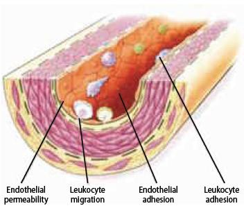
138 KB | Figure 7. Endothelial dysfunction: Leukocyte adhesion and migration into the deep layer of the intima. | 1 |
| 13:00, 10 January 2012 | Figure 8 - Fatty streak formation.png (file) |  |
138 KB | Figure 8. Fatty streak formation | 1 |
| 12:52, 10 January 2012 | Figure 7 - Endothelial dysfunction.png (file) |  |
142 KB | Figure 7. Endothelial dysfunction | 1 |
| 12:47, 13 January 2012 | Figure 1 - algorithm for the initial evaluation of patients with clinical symptoms of angina.png (file) |  |
149 KB | Figure 1 Algorithm for the initial evaluation of patients with clinical symptoms of angina | 1 |
| 16:33, 1 February 2012 | Figure 14. Congenitally corrected transposition of the great arteries.png (file) | 173 KB | {{Information |Description=Figure 14. Congenitally corrected transposition of the great arteries. RA, right atrium. LA, left atrium. RV, right ventricle. LV, left ventricle. p, pulmonary artery. ao, aorta. tric, tricuspid valve. |Source=illustration by dr | 1 | |
| 01:11, 11 January 2012 | Figure 8 - Endothelial dysfunction - Leukocyte adhesion and migration into the deep layer of the intima.png (file) |  |
188 KB | Figure 8. Endothelial dysfunction: Leukocyte adhesion and migration into the deep layer of the intima. | 1 |
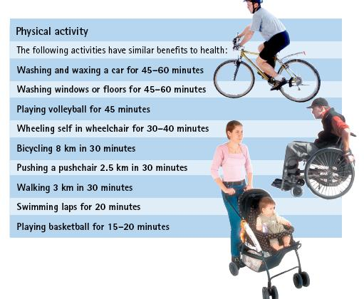
| 13:03, 10 January 2012 | Figure 14 - Recommendations for physical activity.png (file) |  |
212 KB | Figure 14. Recommendations for physical activity | 1 |
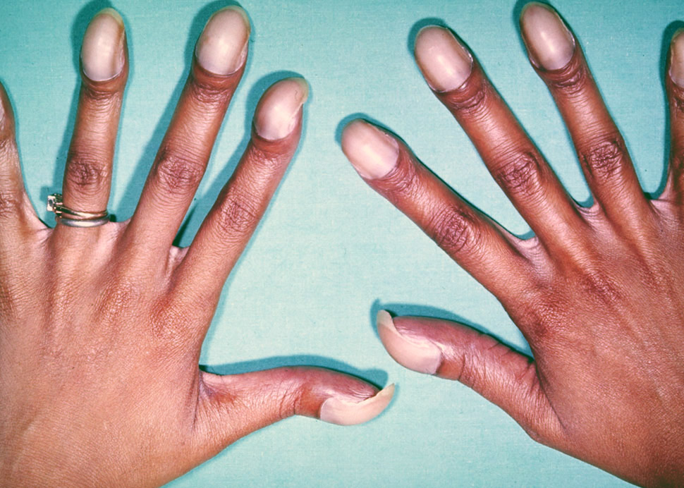
| 15:22, 25 January 2012 | 22. Eisenmenger.jpg (file) |  |
219 KB | {{Information |Description=Figure 22. Photo showing typical features of chronic hypoxemia in Eisenmenger syndrome, with typical digital clubbing with cyanotic nail beds. |Source=from commons.wikipedia.org |Date=Published: |Author= |Permission= |other_vers | 1 |
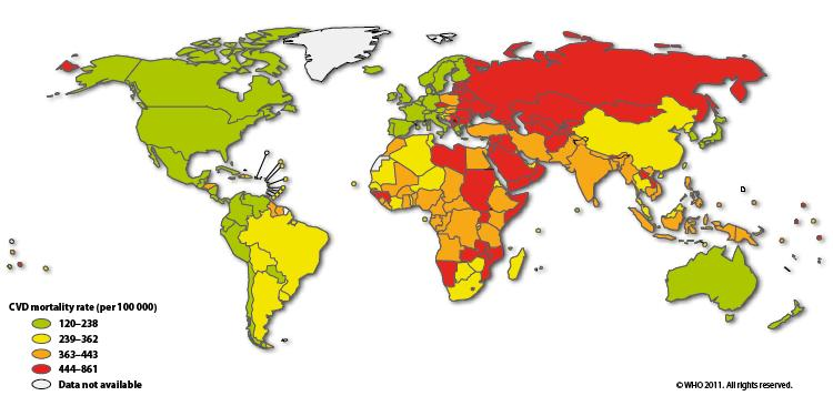
| 12:44, 10 January 2012 | Figure 1 - World map CVD mortality rates in males.png (file) |  |
240 KB | Figure 1. World map CVD mortality rates in males | 1 |
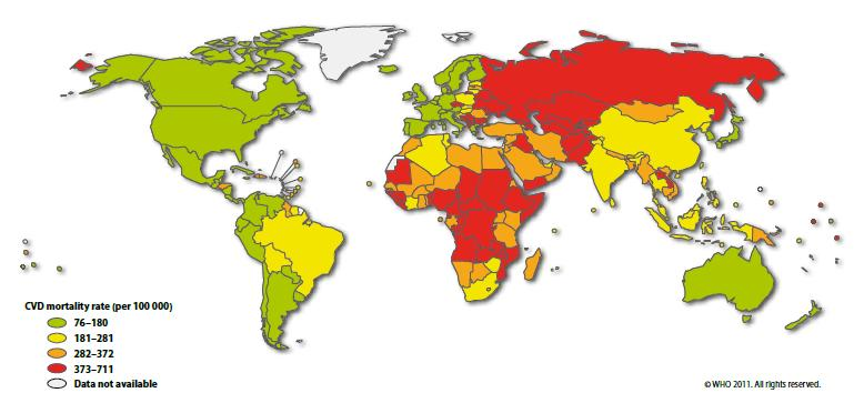
| 12:45, 10 January 2012 | Figure 2 - World map CVD mortality rates in females.png (file) |  |
249 KB | Figure 2. World map CVD mortality rates in females | 1 |
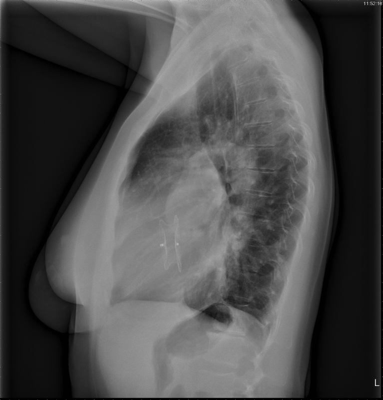
| 23:58, 22 January 2012 | 23. Amplatzer.jpg (file) |  |
268 KB | {{Information |Description=Figure 23. Chest radiograph, left lateral view, of a 34-year old female who recently underwent percutaneous closure of her ASD with an Amplatzer device. |Source=from commons.wikipedia.org |Date=Published: |Author= |Permission= | | 1 |
| 14:55, 25 January 2012 | 24. univentricular heart.PNG (file) |  |
325 KB | {{Information |Description=Figure 24. Echocardiographic image of a male patient with a univentricular heart. |Source=from commons.wikipedia.org |Date=Published: |Author= |Permission= |other_versions= }} | 1 |
| 14:31, 25 January 2012 | 10. coarctatie repair.PNG (file) |  |
394 KB | {{Information |Description=Figure 10. Schematic drawing showing surgical procedures for repair of coarctation of the aorta. Left: resection with end-to-end anastomosis. Middle: dilating technique using a patch; this technique is used in coarctations invol | 1 |
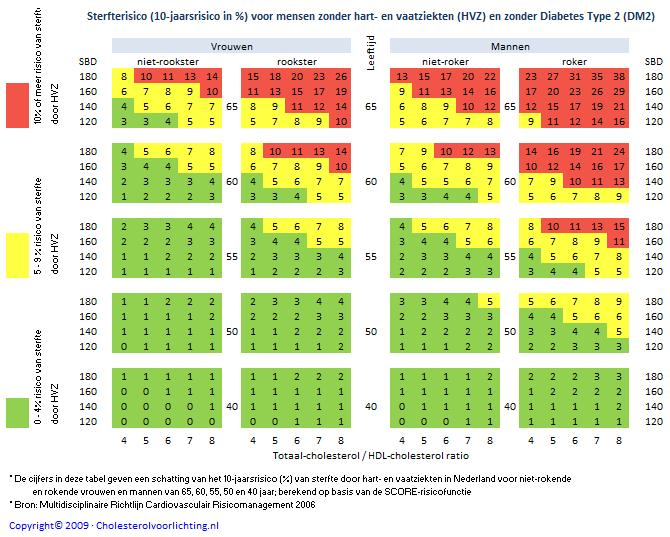
| 15:09, 13 January 2012 | Mortality risk for people with heart disease and type 2 diabetes.png (file) |  |
432 KB | Mortality Risk (10-year risk in %) for People with Heart Disease (HVZ) and Type 2 Diabetes (DM2) | 1 |
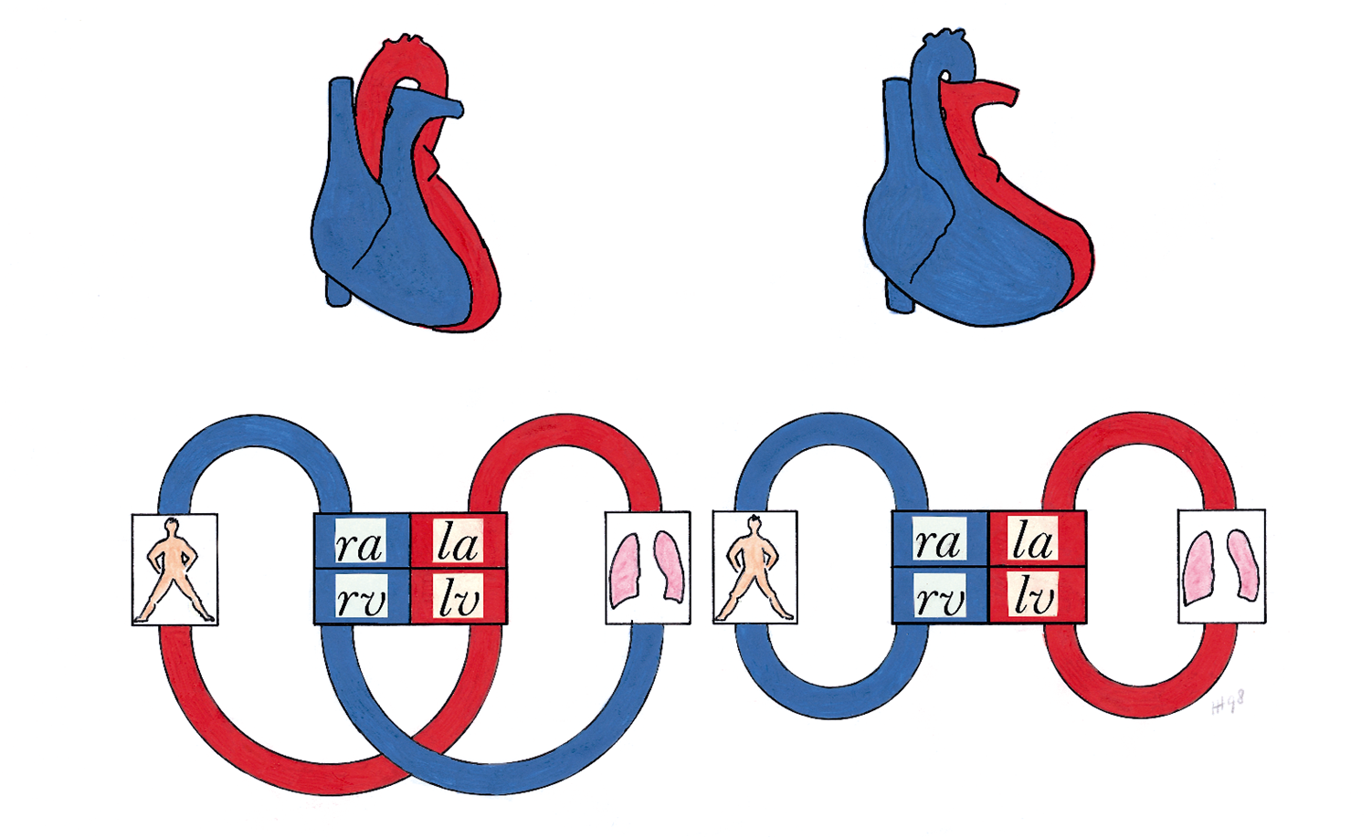
| 14:42, 25 January 2012 | 13. TGA.PNG (file) |  |
492 KB | {{Information |Description=Figure 13. Schematic drawing of the circulation in transposition of the great arteries. Left: normal position of the great arteries with the pulmonary and systemic circulation serially connected. Right: transposition of the grea | 1 |
| 15:19, 25 January 2012 | 21. Ebstein.PNG (file) |  |
495 KB | {{Information |Description=Figure 21. Schematic drawing showing Ebstein’s anomaly of the tricuspid valve. Left: normal heart with openend right ventricle. Right: Ebstein’s anomaly with displacement of the septal and posterior tricuspid leaflet, leadin | 1 |
| 16:31, 14 January 2012 | SaddlePE.PNG (file) |  |
513 KB | Chest spiral CT scan with radiocontrast agent showing multiple filling defects both at the bifurcation and in the pulmonary arteries. | 1 |
| 14:30, 25 January 2012 | 9. coarctatie.PNG (file) |  |
594 KB | {{Information |Description=Figure 9. Schematic drawing of the anatomy prenatal (left) and postnatal (right) in coarctation of the aorta. In the normal situation (without coarctation) only 10 percent of the fetal cardiac output flows through the descending | 1 |
| 14:32, 25 January 2012 | 11. coarctatie repair2.PNG (file) |  |
640 KB | {{Information |Description=Figure 11. Schematic drawing showing surgical procedures for repair of a coarctation of the aorta. Left: an interposition graft. Middle: the extended aortic arch repair. Right: the extra-anatomical bypass. |Source=commons.wikipe | 1 |
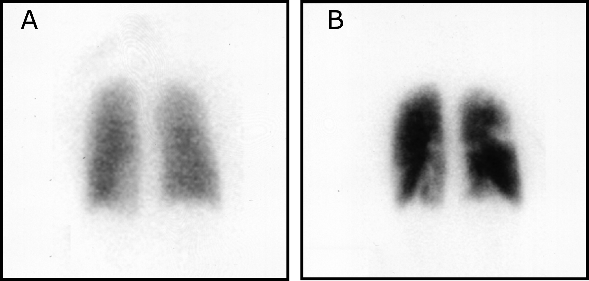
| 16:33, 14 January 2012 | Pulmonary embolism scintigraphy PLoS.png (file) |  |
766 KB | Ventilation-perfusion scintigraphy in a woman taking hormonal contraceptives and valdecoxib. (A) After inhalation of 20.1 mCi of Xenon-133 gas, scintigraphic images were obtained in the posterior projection, showing uniform ventilation to lungs. (B) After | 1 |
| 16:31, 1 February 2012 | Figure 13. Schematic drawing of the circulation in transposition of the great arteries.png (file) | 821 KB | {{Information |Description=Figure 13. Schematic drawing of the circulation in transposition of the great arteries. Left: normal position of the great arteries with the pulmonary and systemic circulation serially connected. Right: transposition of the grea | 1 | |
| 16:19, 1 February 2012 | Figure 10. Schematic drawing showing surgical procedures for repair of coarctation of the aorta.png (file) |  |
1.22 MB | {{Information |Description=Figure 10. Schematic drawing showing surgical procedures for repair of coarctation of the aorta. Left: resection with end-to-end anastomosis. Middle: dilating technique using a patch; this technique is used in coarctations invol | 1 |