Uploads by NiloferT
Jump to navigation
Jump to search
This special page shows all uploaded files.
| Date | Name | Thumbnail | Size | Description | Versions |
|---|---|---|---|---|---|
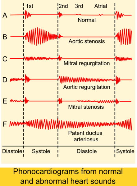
| 18:27, 28 November 2012 | 441px-Phonocardiograms from normal and abnormal heart sounds.png (file) | 125 KB | Description: Phonocardiograms from normal and abnormal heart sounds Author: Madhero88 Source:http://en.wikipedia.org/wiki/File:Phonocardiograms_from_normal_and_abnormal_heart_sounds.png | 1 | |
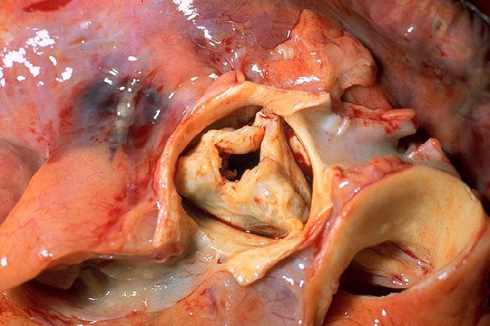
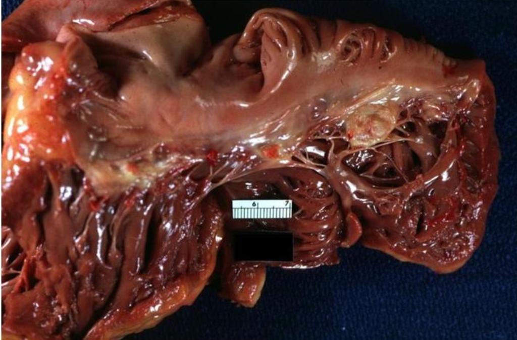
| 18:28, 9 October 2012 | Aortic stenosis rheumatic, gross pathology 20G0014 lores.jpg (file) |  |
92 KB | Description: Gross pathology of rheumatic heart disease: aortic stenosis. Aorta has been removed to show thickened, fused aortic valve leaflets and opened coronary arteries from above. Autopsy. Content Providers(s). Author: CDC/Dr. Edwin P. Ewing, Jr. ... | 1 |
| 20:23, 9 October 2012 | Aortic valve (1).gif (file) | .gif) |
2.23 MB | This is a video clip from a living, beating pig heart that was prepared in the laboratory as a working Langendorf preparation. The heart was arrested, connected to the perfusion system and restarted. The working fluid was oxygenated balanced saline sol... | 2 |
| 18:38, 18 October 2012 | Apical 4 chamber view.gif (file) |  |
72 KB | Description: Apical four chamber view of heart Date: 10 July 1999 Source: http://www.yale.edu/imaging/echo_atlas/views/four_chamber.html Author: Patrick J. Lynch and C. Carl Jaffe | 1 |
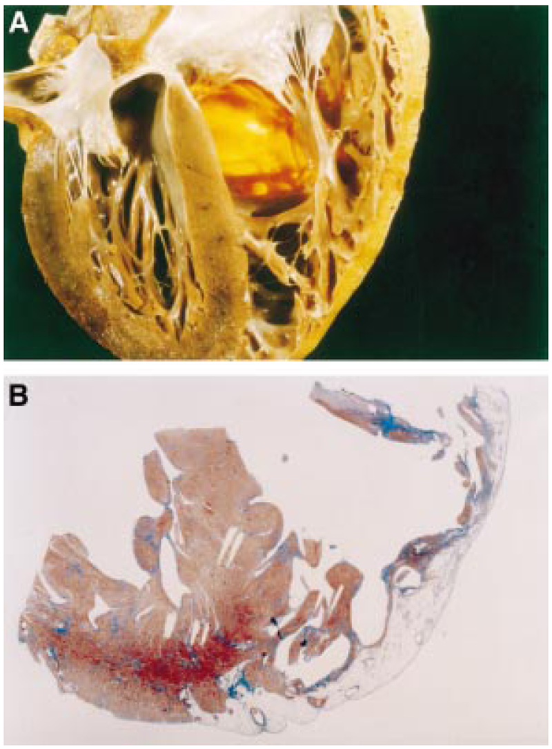
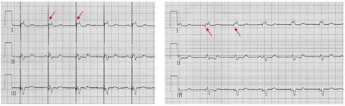
| 21:50, 31 October 2012 | Arvdhart.png (file) |  |
964 KB | 1 | |
| 04:41, 6 August 2012 | AtrialCap.jpg (file) |  |
75 KB | 1 | |
| 19:19, 6 July 2012 | Bb reentry small.svg (file) | 52 KB | 5 | ||
| 23:41, 23 December 2012 | Blue circle for diabetes.svg.png (file) |  |
8 KB | Description: The blue circle is the global symbol for diabetes, introduced by the International Diabetes Federation with the aim of giving diabetes a common identity, supporting existing efforts to raise awareness of diabetes and placing the diabetes e... | 1 |
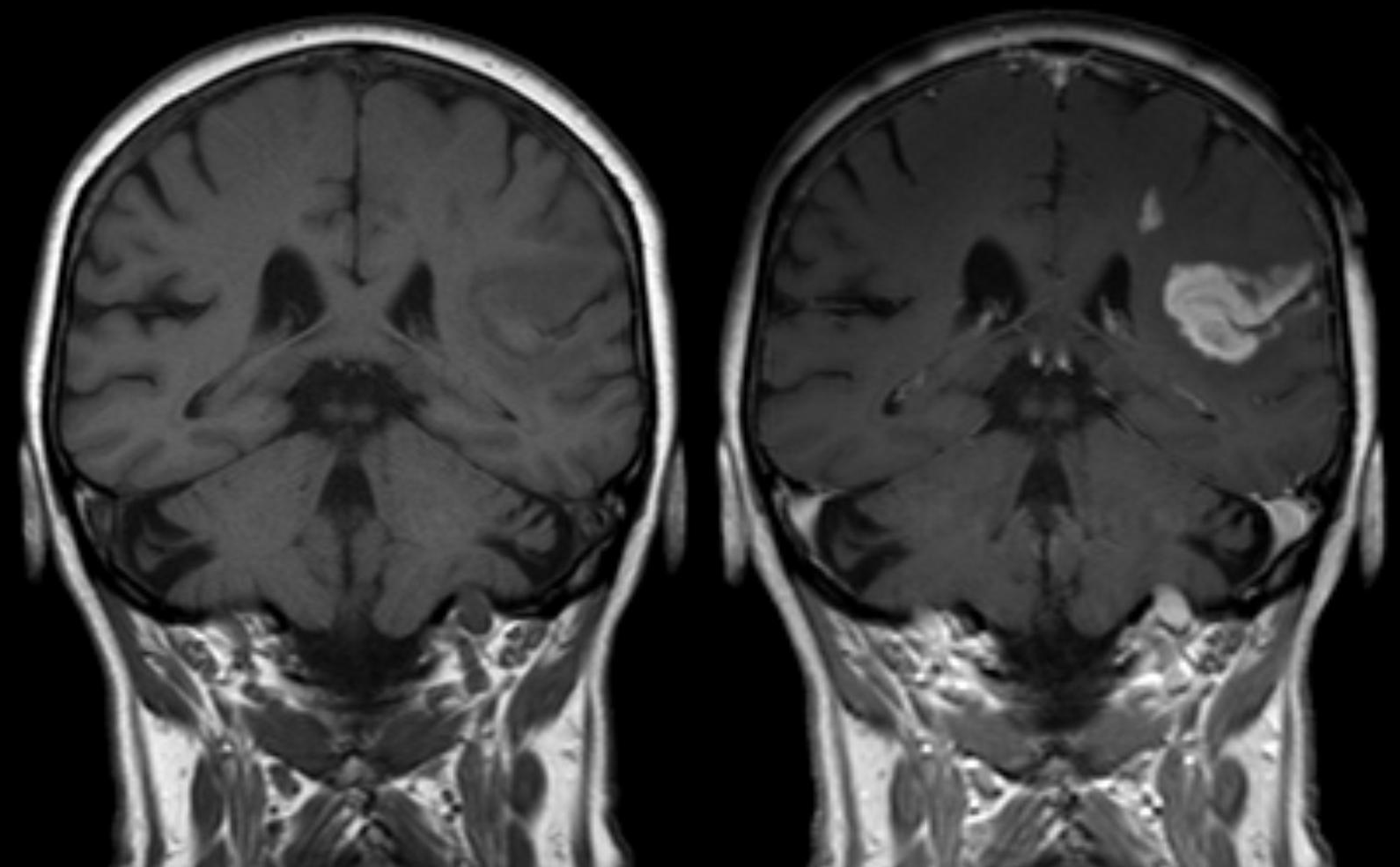
| 14:21, 17 January 2013 | Bluthirnschranke nach Infarkt nativ und KM.png (file) |  |
549 KB | Description: Defect of the blood-brain barrier after stroke shown in MRI. T1-weighted images, left image without right image with contrast medium administration. Deutsch: Störung der Blut-Hirn-Schranke nach einem ischämischen Hirninfarkt im Stromgeb... | 1 |
| 09:21, 9 October 2012 | Brugada.jpg (file) |  |
13 KB | 1 | |
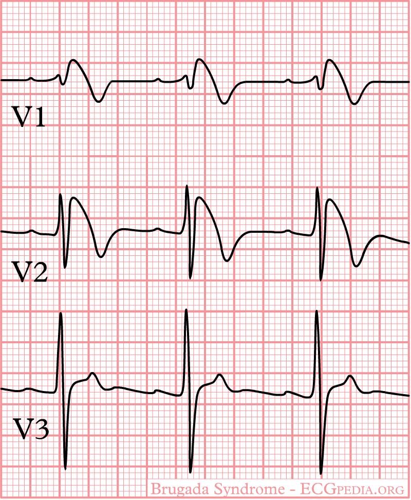
| 09:21, 9 October 2012 | Brugada.png (file) |  |
29 KB | 1 | |
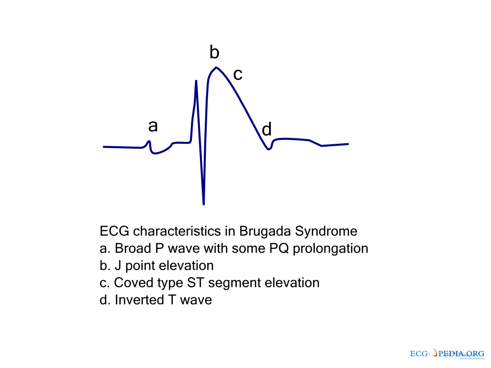
| 09:21, 9 October 2012 | Brugada ecg characteristics.png (file) |  |
39 KB | 1 | |
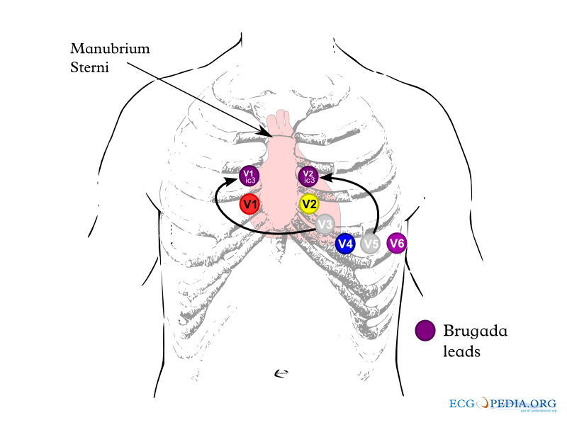
| 09:21, 9 October 2012 | Brugada lead placement.png (file) |  |
84 KB | 1 | |
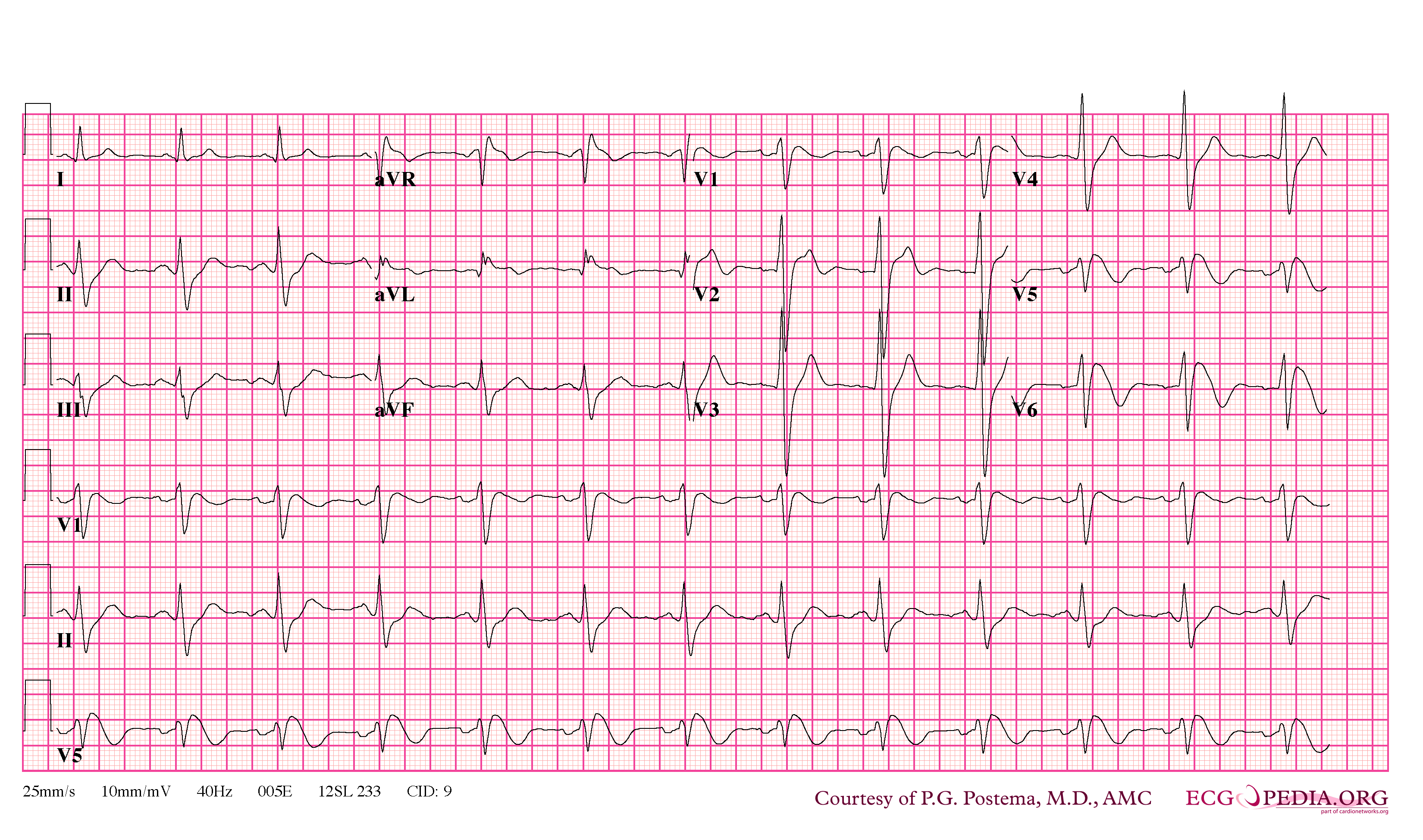
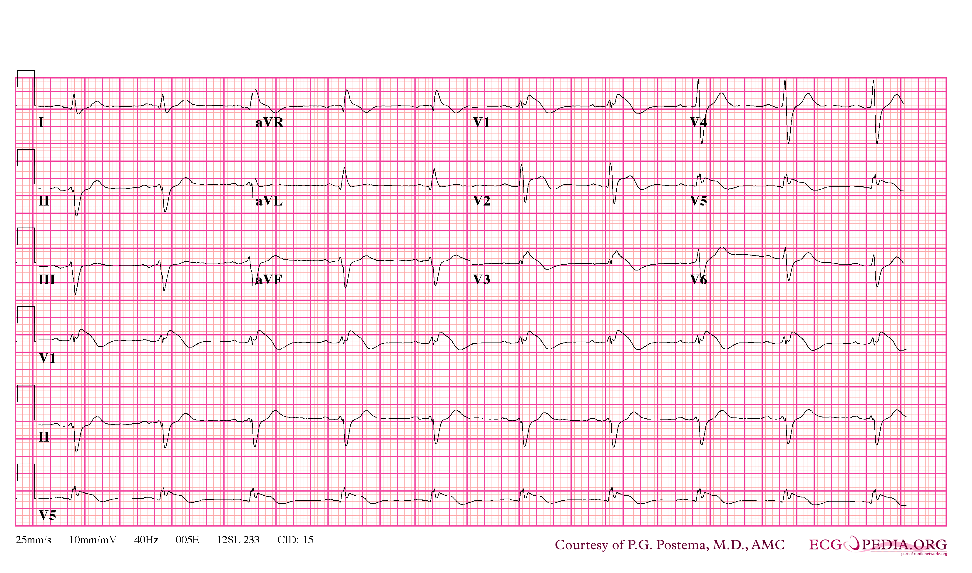
| 09:25, 9 October 2012 | Brugada syndrome type1 example1.png (file) |  |
370 KB | 1 | |
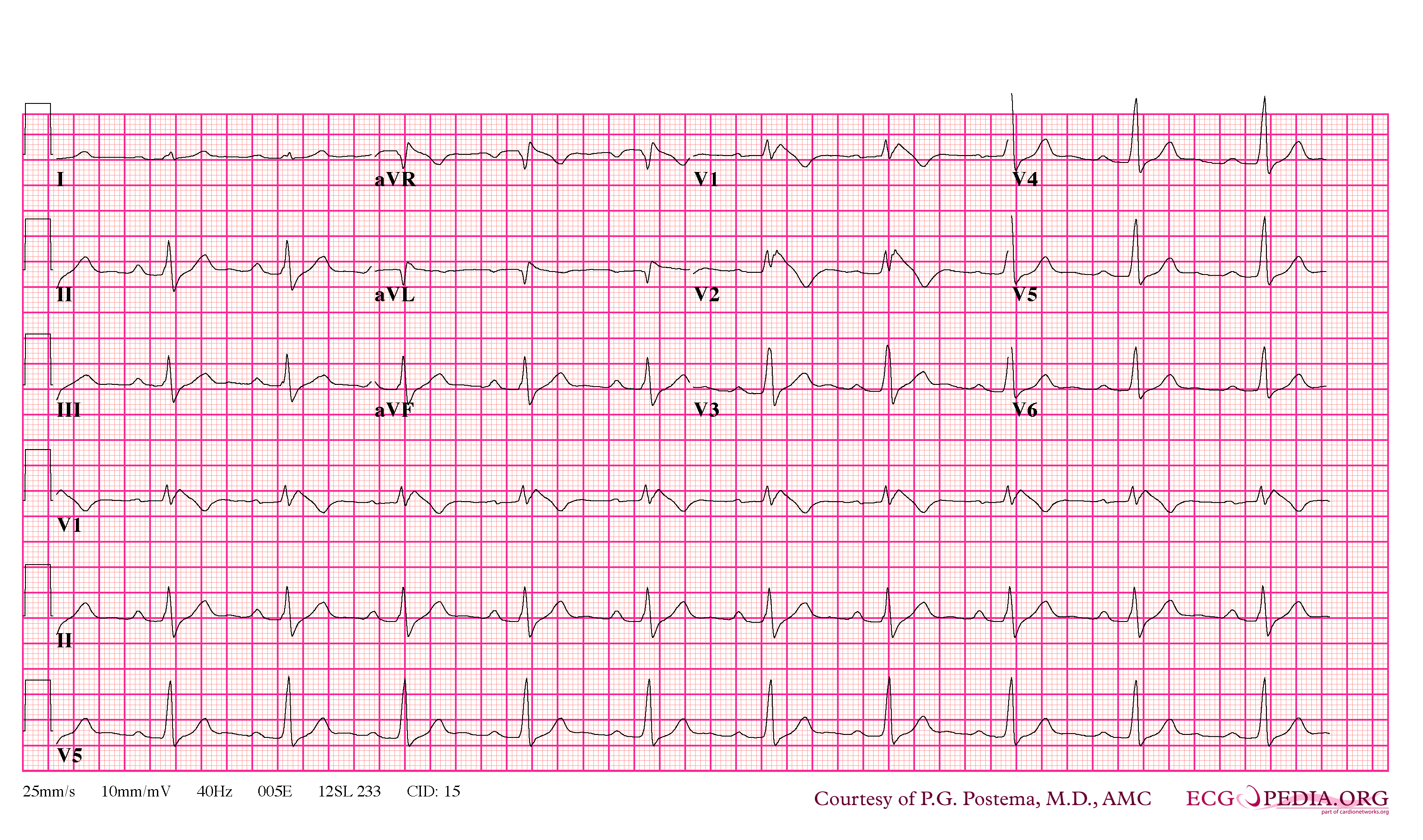
| 09:26, 9 October 2012 | Brugada syndrome type1 example2.png (file) |  |
348 KB | 1 | |
| 09:22, 9 October 2012 | Brugada syndrome type1 example3.png (file) |  |
63 KB | 1 | |
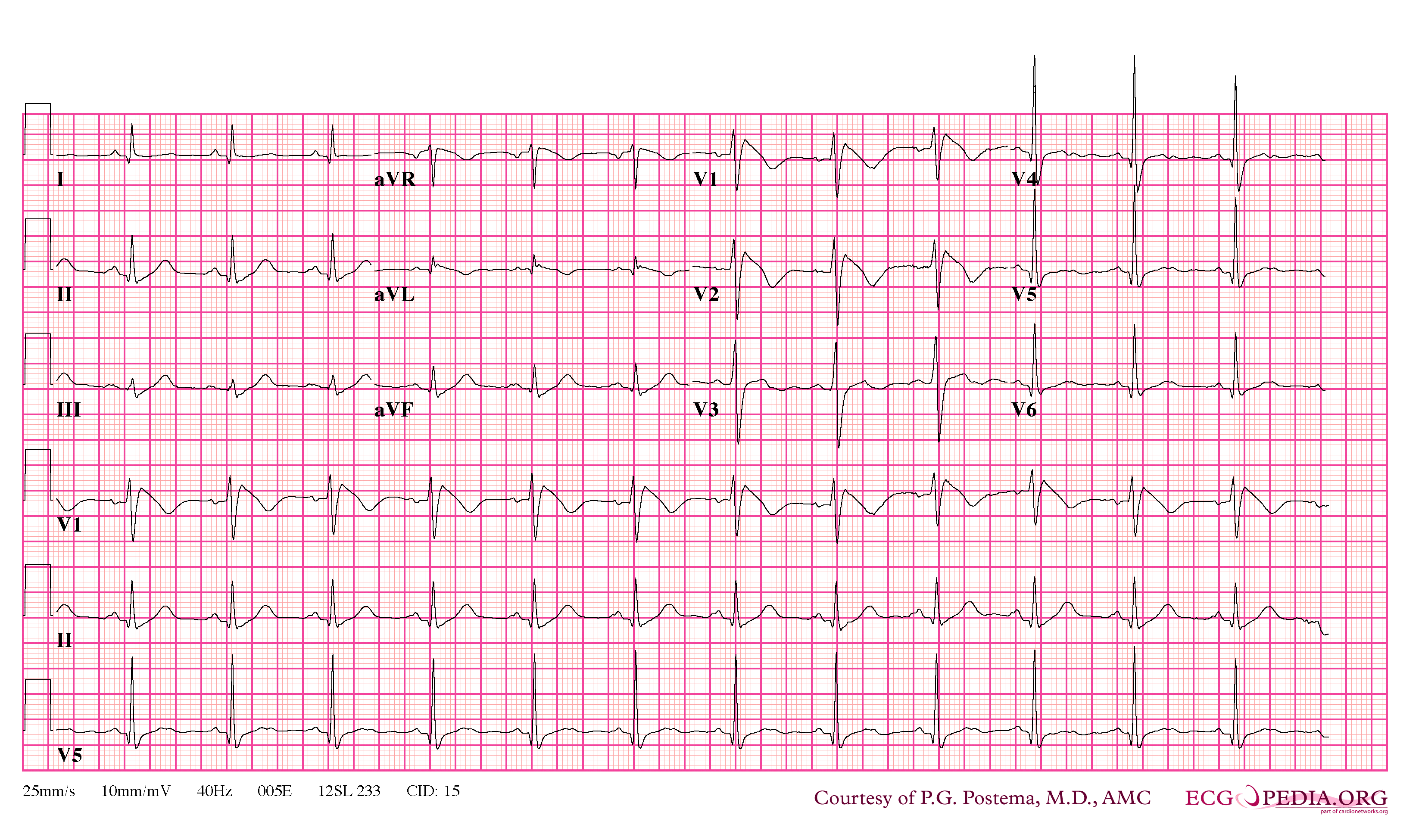
| 09:24, 9 October 2012 | Brugada syndrome type1 example4.png (file) |  |
364 KB | 1 | |
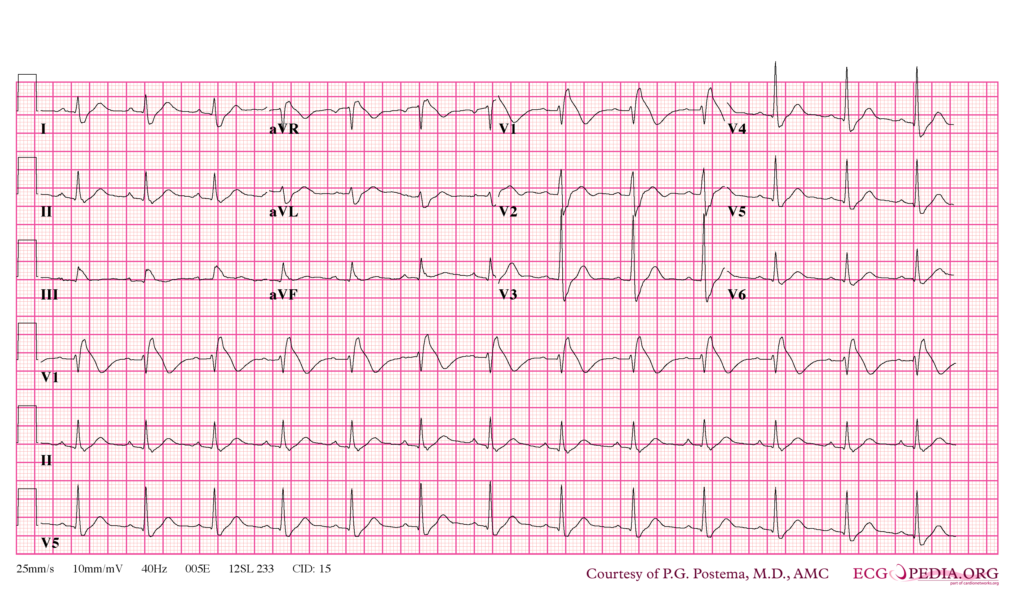
| 09:25, 9 October 2012 | Brugada syndrome type1 example5.png (file) |  |
364 KB | 1 | |
| 09:31, 9 October 2012 | Brugada syndrome type1 example6.jpg (file) |  |
970 KB | 1 | |
| 09:26, 9 October 2012 | Brugada syndrome type2 example1.png (file) |  |
363 KB | 1 | |
| 09:29, 9 October 2012 | Brugada syndrome type2 example2.jpg (file) |  |
677 KB | 1 | |
| 19:52, 5 August 2012 | ChestXray.jpg (file) |  |
46 KB | 2 | |
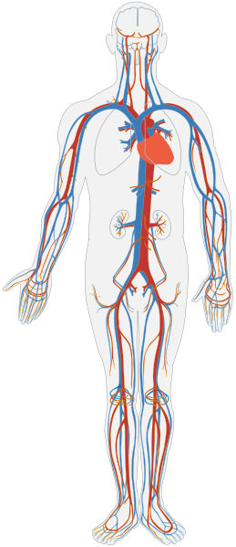
| 18:42, 29 November 2012 | Circulatory System no tags.svg (file) |  |
104 KB | 2 | |
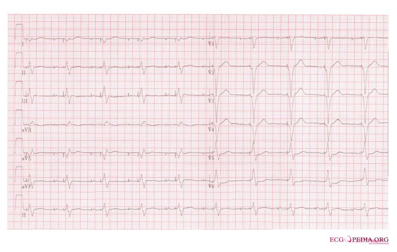
| 00:20, 6 August 2012 | Ddd paced 12lead.jpg (file) |  |
73 KB | 1 | |
| 22:52, 23 December 2012 | Diabetes County level estimates 2004-2009.gif (file) |  |
363 KB | Description: An animated map of the United States showing the prevalence of diabetes from 2004-2009. Date: 12 August 2012 Source: http://www.cdc.gov/obesity/data/adult.html from http://apps.nccd.cdc.gov/DDT_STRS2/NationalDiabetesPrevalenceEstimates.asp... | 1 |
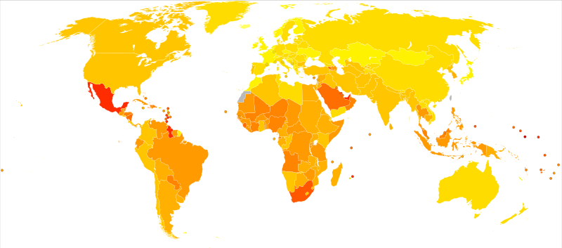
| 23:11, 23 December 2012 | Diabetes mellitus world map - DALY - WHO2004.svg (file) |  |
78 KB | Description: Age-standardised disability-adjusted life year (DALY) rates from Diabetes mellitus by country (per 100,000 inhabitants). Date: 11 January 2010 Source: Vector map from BlankMap-World6, compact.svg by Canuckguy et al. Data from Death and DAL... | 1 |
| 00:24, 4 January 2013 | Diabetes world map - 2000.png (file) |  |
1.45 MB | 2 | |
| 18:23, 9 October 2012 | Diagram of the human heart (valves improved).svg (file) | .svg) |
27 KB | Source:http://commons.wikimedia.org/wiki/Image:Diagram_of_the_human_heart_%28cropped%29.svg | 2 |
| 03:46, 6 August 2012 | ECGT.jpg (file) |  |
57 KB | 1 | |
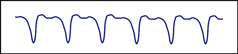
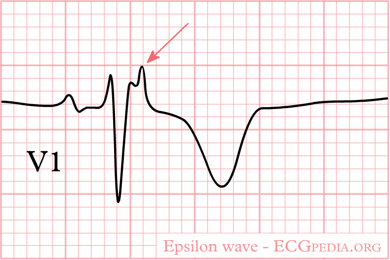
| 21:45, 31 October 2012 | Epsilon wave.png (file) |  |
14 KB | 1 | |
| 10:21, 18 May 2012 | Figure1.jpg (file) |  |
1.06 MB | 1 | |
| 11:25, 18 May 2012 | Figure 10.jpg (file) |  |
1.3 MB | 1 | |
| 11:30, 18 May 2012 | Figure 11.jpg (file) |  |
1.43 MB | 1 | |
| 11:49, 18 May 2012 | Figure 12.jpg (file) |  |
1.99 MB | 1 | |
| 12:48, 20 May 2012 | Figure 13.jpg (file) |  |
1.48 MB | 1 | |
| 10:36, 18 May 2012 | Figure 2.jpg (file) |  |
1.25 MB | 1 | |
| 10:40, 18 May 2012 | Figure 3.jpg (file) |  |
691 KB | 1 | |
| 10:46, 18 May 2012 | Figure 4.jpg (file) |  |
566 KB | 1 | |
| 12:43, 20 May 2012 | Figure 5.jpg (file) |  |
815 KB | 1 | |
| 10:50, 18 May 2012 | Figure 6.jpg (file) |  |
601 KB | 1 | |
| 15:25, 20 May 2012 | Figure 7.jpg (file) |  |
1.47 MB | Reverted to version as of 15:01, 20 May 2012 | 5 |
| 11:10, 18 May 2012 | Figure 8.jpg (file) |  |
1.27 MB | 1 | |
| 11:35, 18 May 2012 | Figure 9.jpg (file) |  |
1.19 MB | 2 | |
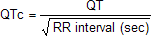
| 23:31, 17 October 2012 | Formule QTc.png (file) | 754 bytes | 1 | ||
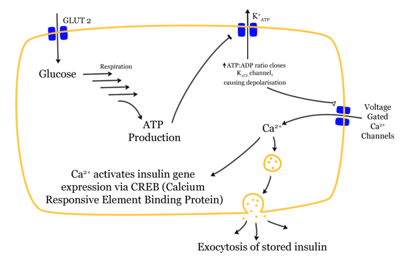
| 22:07, 23 December 2012 | Glucose-insulin-release.png (file) |  |
76 KB | Description: Mechanism of glucose dependent insulin release Date:16 August 2004 (original upload date) Source:Transferred from en.wikipedia Author:Prisonblues | 1 |
| 02:00, 14 July 2012 | Graph.jpg (file) |  |
52 KB | 1 | |
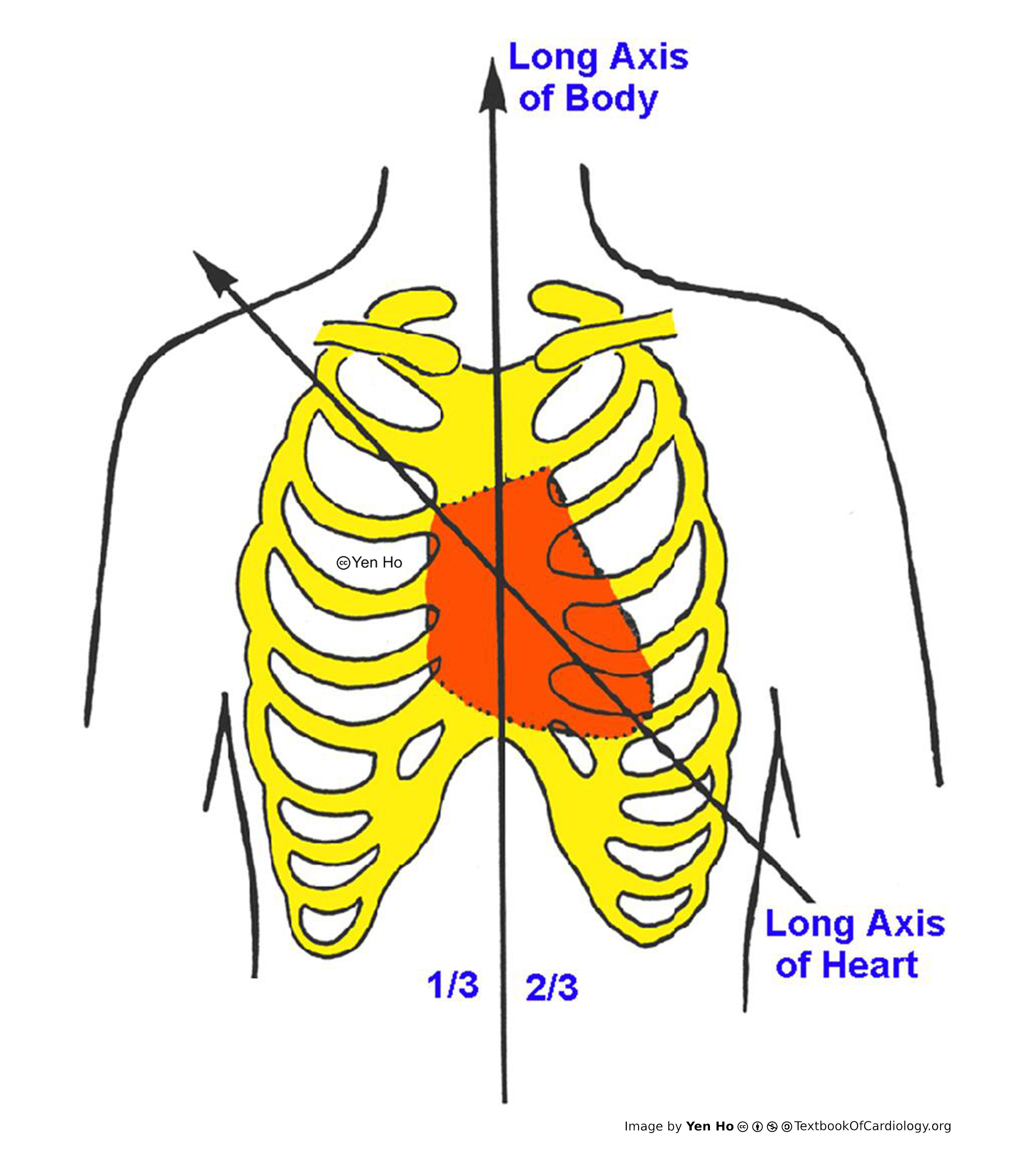
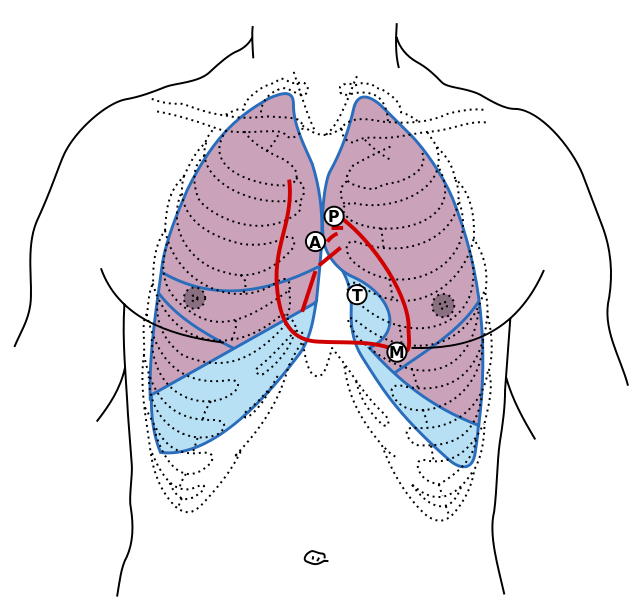
| 15:29, 28 November 2012 | Gray1216 modern locations.svg.png (file) |  |
109 KB | Description: Front of thorax, showing surface relations of bones, lungs (purple), pleura (blue), and heart (red outline). Heart valves are labeled (Mitral (Bicuspid), Tricuspid, Aortic, Pulmonary). Figure 1216 from Gray's Anatomy. It has been updated t... | 1 |
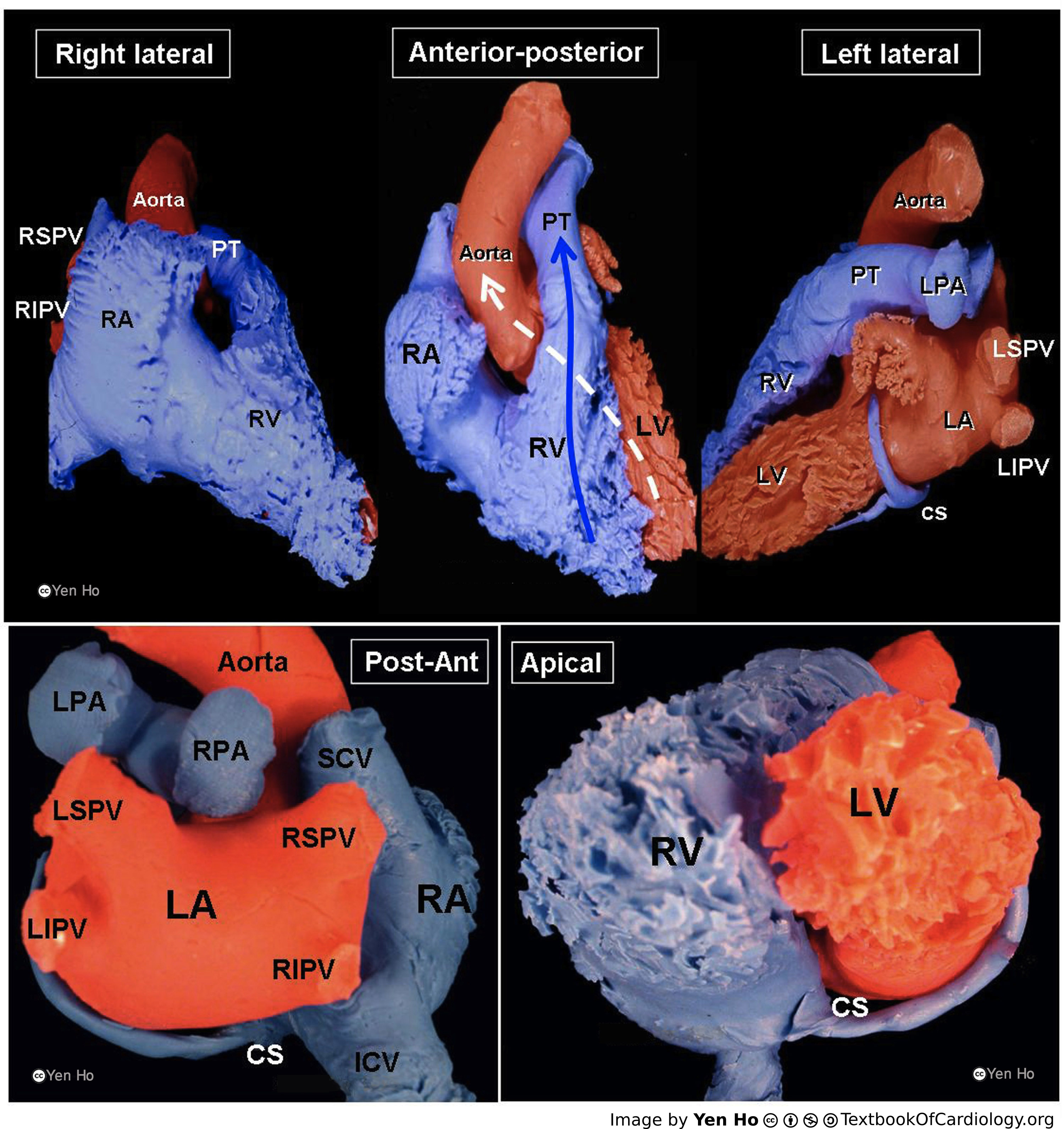
| 04:43, 28 November 2013 | Heart1.JPG (file) |  |
81 KB | 1 | |
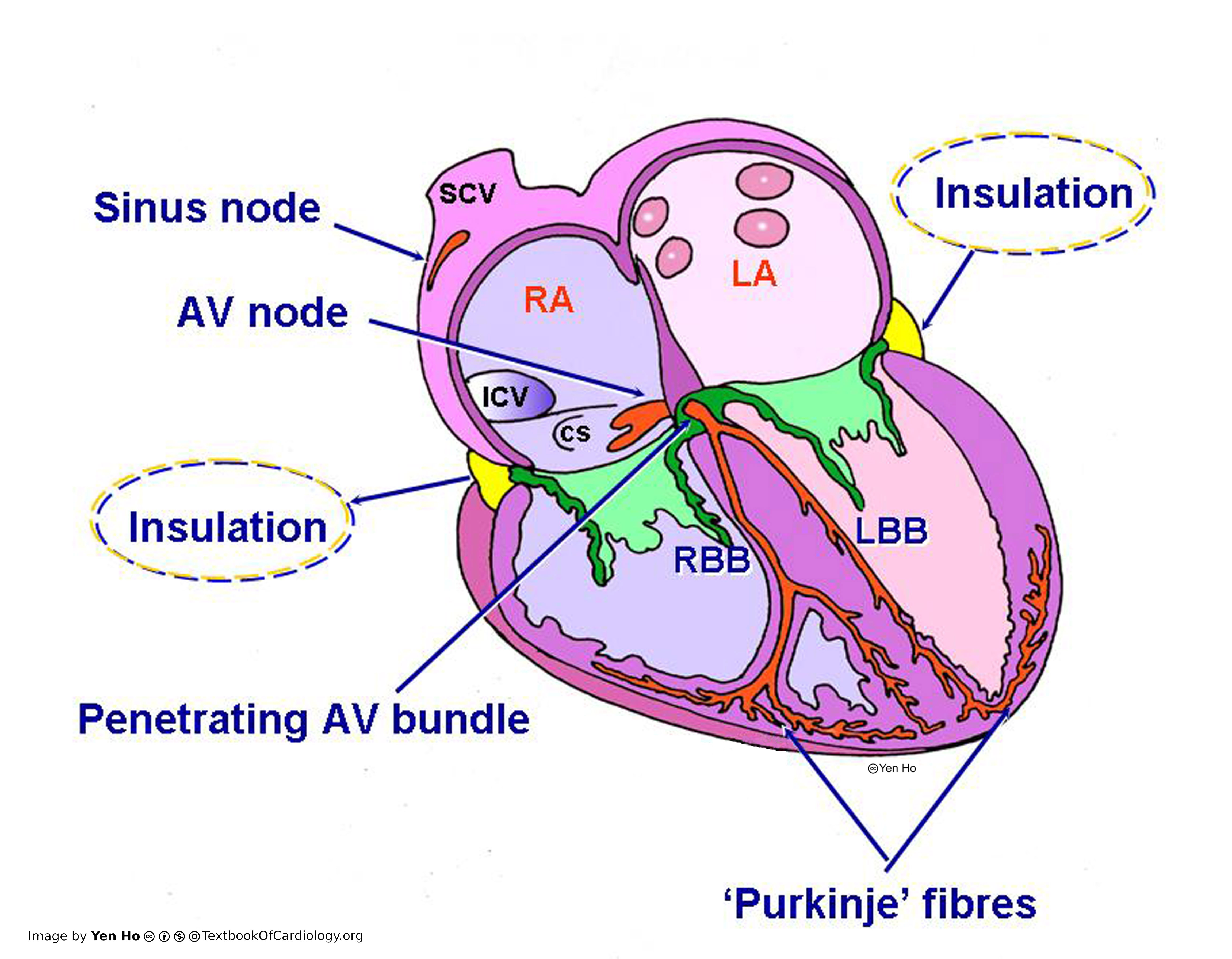
| 04:45, 28 November 2013 | Heart2.JPG (file) |  |
84 KB | 1 | |
| 04:46, 28 November 2013 | Heart3.JPG (file) |  |
65 KB | 1 |