Uploads by NiloferT
This special page shows all uploaded files.
| Date | Name | Thumbnail | Size | Description | Versions |
|---|---|---|---|---|---|
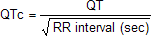
| 23:31, 17 October 2012 | Formule QTc.png (file) | 754 bytes | 1 | ||
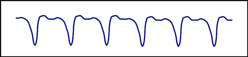
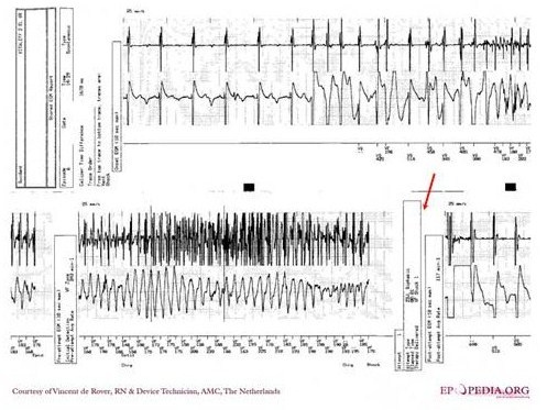
| 00:14, 6 August 2012 | Paced2.gif (file) |  |
7 KB | Reverted to version as of 23:31, 5 August 2012 | 3 |
| 23:41, 23 December 2012 | Blue circle for diabetes.svg.png (file) |  |
8 KB | Description: The blue circle is the global symbol for diabetes, introduced by the International Diabetes Federation with the aim of giving diabetes a common identity, supporting existing efforts to raise awareness of diabetes and placing the diabetes e... | 1 |
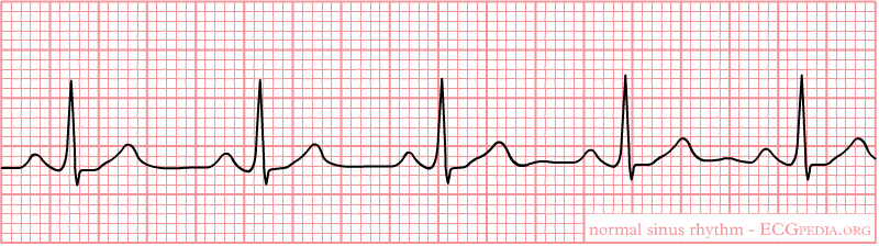
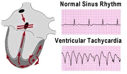
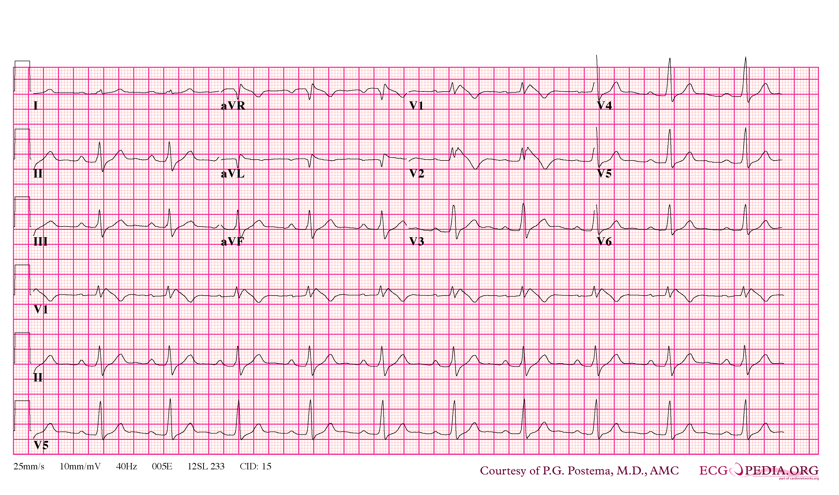
| 23:31, 17 October 2012 | Nsr.png (file) |  |
11 KB | 1 | |
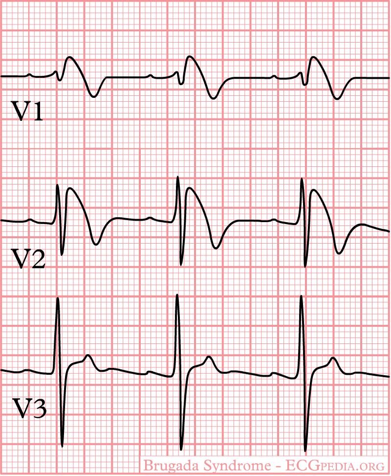
| 09:21, 9 October 2012 | Brugada.jpg (file) |  |
13 KB | 1 | |
| 18:41, 29 November 2012 | P1.jpg (file) |  |
13 KB | 1 | |
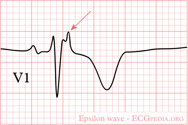
| 21:45, 31 October 2012 | Epsilon wave.png (file) |  |
14 KB | 1 | |
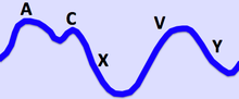
| 14:56, 28 November 2012 | Jugular Venous Pulse.png (file) |  |
16 KB | Description: The Jugular Venous Pressure Waveform Date:13 December 2011 Author Ecgtocardiology | 1 |
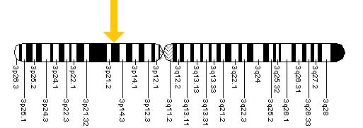
| 09:25, 9 October 2012 | Scn5a.jpg (file) |  |
17 KB | 1 | |
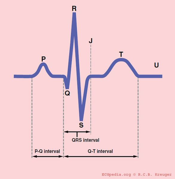
| 18:47, 18 October 2012 | QRSwaves.jpg (file) |  |
20 KB | Description: The different waves and intervals of the ECG (P, PQ, QRS, QT, ST) Date: 2007 Author: Rob Kreuger, medical illustrator, AMC, The Netherlands | 1 |
| 04:52, 1 July 2012 | Orthostatic.JPG (file) |  |
25 KB | 1 | |
| 18:23, 9 October 2012 | Diagram of the human heart (valves improved).svg (file) | .svg) |
27 KB | Source:http://commons.wikimedia.org/wiki/Image:Diagram_of_the_human_heart_%28cropped%29.svg | 2 |
| 18:29, 9 October 2012 | Heart bicuspid aortic valve.svg (file) |  |
28 KB | Description: Heart bicuspid aortic valve anatomy Date: 23 December 2006 Author: Patrick J. Lynch, medical illustrator Creative Commons Attribution 2.5 License 2006 | 1 |
| 09:21, 9 October 2012 | Brugada.png (file) |  |
29 KB | 1 | |
| 04:47, 1 July 2012 | Pathophysiology.JPG (file) |  |
30 KB | 1 | |
| 03:35, 6 August 2012 | RedTh.jpg (file) | 31 KB | 1 | ||
| 19:28, 6 July 2012 | Vent1.jpg (file) |  |
39 KB | 1 | |
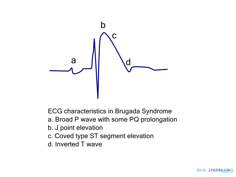
| 09:21, 9 October 2012 | Brugada ecg characteristics.png (file) |  |
39 KB | 1 | |
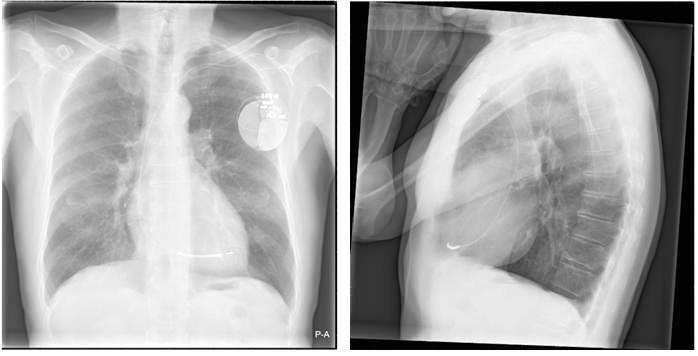
| 20:14, 5 August 2012 | Xthorax.jpg (file) |  |
44 KB | 1 | |
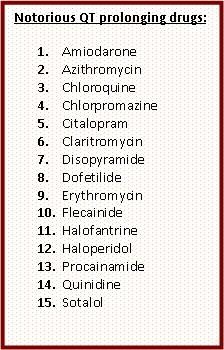
| 20:32, 6 July 2012 | VentDrg.jpg (file) |  |
45 KB | 1 | |
| 19:52, 5 August 2012 | ChestXray.jpg (file) |  |
46 KB | 2 | |
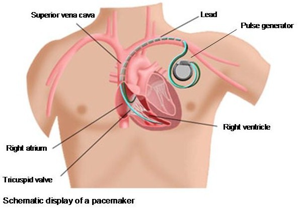
| 17:54, 5 August 2012 | SchematicImg.jpg (file) |  |
50 KB | 2 | |
| 19:19, 6 July 2012 | Bb reentry small.svg (file) | 52 KB | 5 | ||
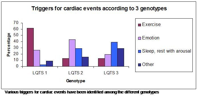
| 02:00, 14 July 2012 | Graph.jpg (file) |  |
52 KB | 1 | |
| 22:47, 5 August 2012 | Schematicpic.jpg (file) |  |
53 KB | 2 | |
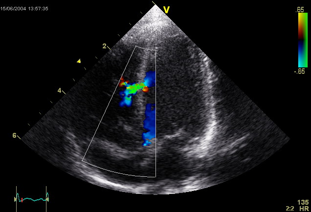
| 23:48, 17 October 2012 | Ventricular Septal Defect.jpg (file) |  |
54 KB | This is an ultrasound picture of the heart, an echocardiogram. It depicts a ventricular septal defect. Author: Kjetil Lenes. | 1 |
| 22:23, 5 August 2012 | Schematic.jpg (file) |  |
54 KB | 1 | |
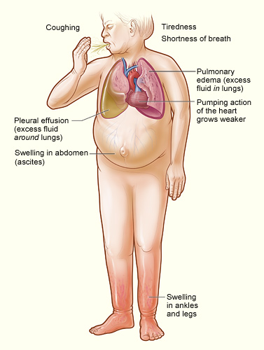
| 13:41, 17 January 2013 | Heartfailure.jpg (file) |  |
55 KB | Description: The illustration shows the major signs and symptoms of heart failure. Date: Originally uploaded to en.wikipedia on 22 September 2008. Source: http://www.nhlbi.nih.gov/health/dci/Diseases/Hf/HF_SignsAndSymptoms.html; transferred from en.w... | 1 |
| 21:38, 28 November 2013 | Heart4.JPG (file) |  |
56 KB | 1 | |
| 03:46, 6 August 2012 | ECGT.jpg (file) |  |
57 KB | 1 | |
| 03:18, 6 January 2013 | Swe.jpg (file) |  |
57 KB | 3 | |
| 18:30, 9 October 2012 | Pulmonary valve stenosis.svg (file) |  |
59 KB | Description: The diagram shows a healthy heart and one suffering from Pulmonary valve stenosis. Date: 12 June 2006 Author: Mariana Ruiz LadyofHats | 1 |
| 20:04, 4 October 2012 | Myocardi1.jpg (file) |  |
61 KB | 1 | |
| 09:22, 9 October 2012 | Brugada syndrome type1 example3.png (file) |  |
63 KB | 1 | |
| 18:50, 18 October 2012 | LeftVentricleShortAxis.gif (file) |  |
64 KB | Description: Short axis view of left ventricle of heart Date: 10 July 1999 Source:http://www.yale.edu/imaging/echo_atlas/views/short_axis_lv.html Author: Patrick J. Lynch and C. Carl Jaffe | 1 |
| 04:46, 28 November 2013 | Heart3.JPG (file) |  |
65 KB | 1 | |
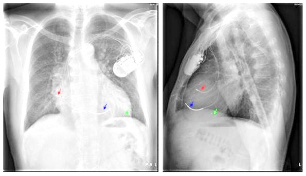
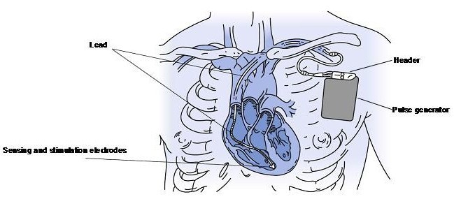
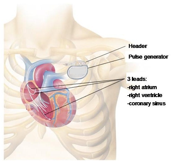
| 02:53, 6 August 2012 | ICD.jpg (file) |  |
67 KB | 1 | |
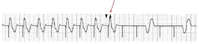
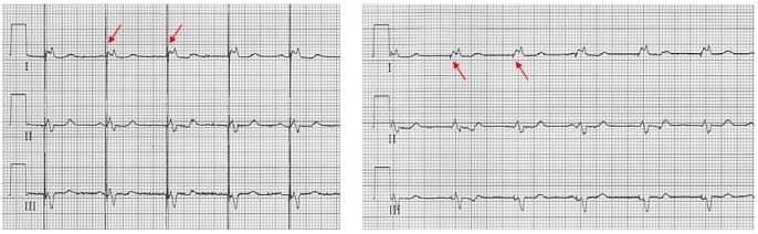
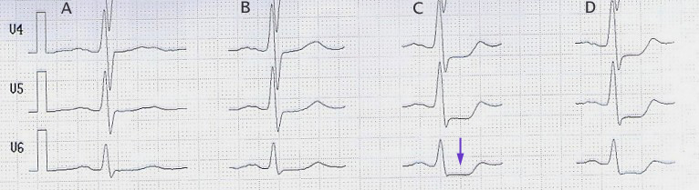
| 23:23, 17 May 2013 | StressECG STDepression.jpg (file) | 72 KB | Description: Belastungs-EKG mit ST-Senkung (Pfeil) ab 100 W (Spalte C) stress-ecg with st-segment-depression (arrow) beginning at 100 W (column C) Date: published 10. Jan. 2006 Source: selbst abgeleitet/own recording Author: J. Heuser JHeuser | 1 | |
| 18:38, 18 October 2012 | Apical 4 chamber view.gif (file) |  |
72 KB | Description: Apical four chamber view of heart Date: 10 July 1999 Source: http://www.yale.edu/imaging/echo_atlas/views/four_chamber.html Author: Patrick J. Lynch and C. Carl Jaffe | 1 |