File:Rheumatic heart disease - 3a - very high mag.jpg
Jump to navigation
Jump to search
Size of this preview: 800 × 533 pixels. Other resolution: 1,280 × 853 pixels.
Original file (1,280 × 853 pixels, file size: 351 KB, MIME type: image/jpeg)
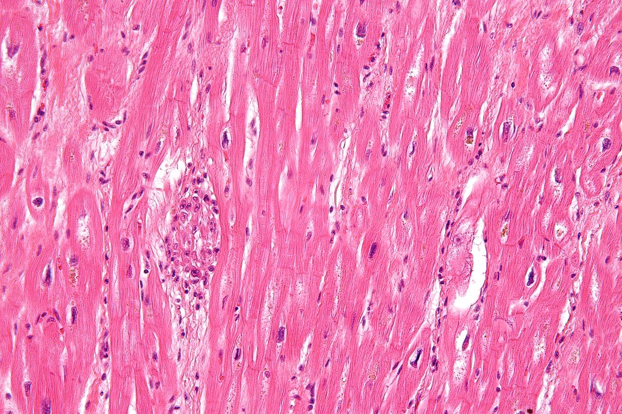
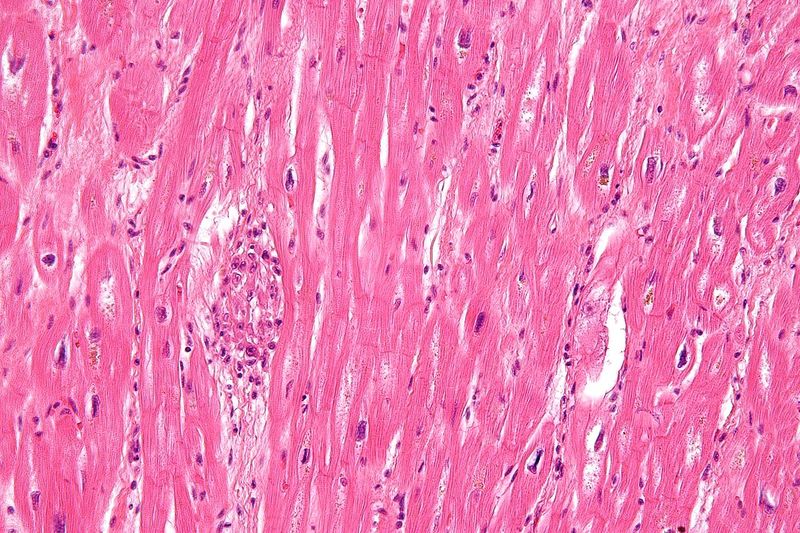
Description: Very high magnification micrograph of rheumatic heart disease. H&E stain. It is due to Streptococcus pyogenes. Microscopic findings include Anitschkow cells (also known as caterpillar cells), and Aschoff bodies. Anitschkow cells are thought to be cardiac histocytes and Aschoff bodies are thought to be granulomas. Author: Nephron
File history
Click on a date/time to view the file as it appeared at that time.
| Date/Time | Thumbnail | Dimensions | User | Comment | |
|---|---|---|---|---|---|
| current | 18:32, 17 May 2013 | | 1,280 × 853 (351 KB) | NiloferT (talk | contribs) | Description: Very high magnification micrograph of rheumatic heart disease. H&E stain. It is due to Streptococcus pyogenes. Microscopic findings include Anitschkow cells (also known as caterpillar cells), and Aschoff bodies. Anitschkow cells are though... |
You cannot overwrite this file.
File usage
The following page uses this file: