File:Pulmonary embolism ECG.jpg: Difference between revisions
Jump to navigation
Jump to search
(Electrocardiogram of a patient with pulmonary embolism showing sinus tachycardia of approximately 150 beats per minute and right bundle branch block.) |
No edit summary |
||
| Line 1: | Line 1: | ||
Electrocardiogram of a patient with pulmonary embolism showing sinus tachycardia of approximately 150 beats per minute and right bundle branch block. | {{Information | ||
|Description={{en|1=Electrocardiogram of a patient with pulmonary embolism showing sinus tachycardia of approximately 150 beats per minute and right bundle branch block.}} | |||
|Source=[http://www.cardiovascularultrasound.com/content/5/1/26 Pulmonary embolism and patent foramen ovale thrombosis: the key role of TEE]. Cardiovascular Ultrasound 2007, 5:26. doi:10.1186/1476-7120-5-26 . Via: commons.wikipedia.org | |||
|Author=Walter Serra, Giuseppe De Iaco, Claudio Reverberi and Tiziano Gherli | |||
|Date=Published: 24 August 2007 | |||
|Permission= | |||
|other_versions= | |||
}} | |||
Latest revision as of 18:48, 22 January 2012
| Description | |
|---|---|
| Source |
Pulmonary embolism and patent foramen ovale thrombosis: the key role of TEE. Cardiovascular Ultrasound 2007, 5:26. doi:10.1186/1476-7120-5-26 . Via: commons.wikipedia.org |
| Date |
Published: 24 August 2007 |
| Author |
Walter Serra, Giuseppe De Iaco, Claudio Reverberi and Tiziano Gherli |
| Permission |
File history
Click on a date/time to view the file as it appeared at that time.
| Date/Time | Thumbnail | Dimensions | User | Comment | |
|---|---|---|---|---|---|
| current | 16:34, 14 January 2012 | 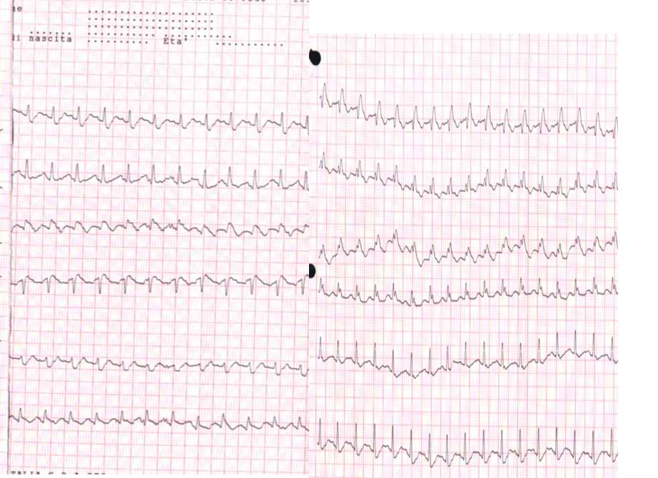 | 922 × 670 (98 KB) | Nja (talk | contribs) | Electrocardiogram of a patient with pulmonary embolism showing sinus tachycardia of approximately 150 beats per minute and right bundle branch block. |
You cannot overwrite this file.
File usage
The following page uses this file: