File:3. VSD.jpg
Jump to navigation
Jump to search
No higher resolution available.
3._VSD.jpg (490 × 298 pixels, file size: 97 KB, MIME type: image/jpeg)
| Description |
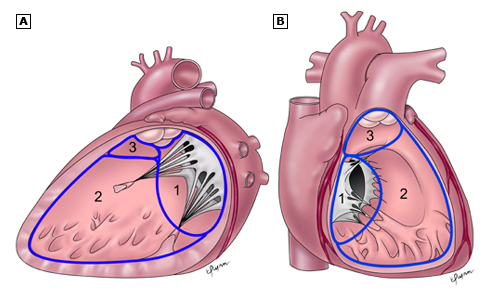
Figure 3. Schematic drawing showing three main anatomic components of the interventricular septum: the septum of the atrioventricular canal (1), the muscular septum (2), the parietal band or distal conal septum (3). |
|---|---|
| Source |
(wordt nagetekend) bron: UptoDate |
| Date | |
| Author | |
| Permission |
File history
Click on a date/time to view the file as it appeared at that time.
| Date/Time | Thumbnail | Dimensions | User | Comment | |
|---|---|---|---|---|---|
| current | 14:48, 25 January 2012 | | 490 × 298 (97 KB) | Nja (talk | contribs) | {{Information |Description=Figure 3. Schematic drawing showing three main anatomic components of the interventricular septum: the septum of the atrioventricular canal (1), the muscular septum (2), the parietal band or distal conal septum (3). |Source=from |
You cannot overwrite this file.
File usage
The following page uses this file: