File:2. ASD2.png: Difference between revisions
Jump to navigation
Jump to search
No edit summary |
No edit summary |
||
| Line 4: | Line 4: | ||
|Date= | |Date= | ||
|Author= Kjetil Lenes | |Author= Kjetil Lenes | ||
|Permission | |Permission=Creative Commons Attribution/Share-Alike License | ||
|other_versions= | |||
}} | }} | ||
Latest revision as of 16:02, 1 February 2012
| Description |
Schematic drawing showing the location of different types of ASD, the view is into an opened right atrium. VCS, superior caval vein. VCI, inferior caval vein. HV, right ventricle. 1, upper sinus venosus defect. 2, lower sinus venosus defect. 3, secundum defect. 4, defect involving coronary sinus. 5, primum defect. |
|---|---|
| Source |
from commons.wikipedia.org |
| Date | |
| Author |
Kjetil Lenes |
| Permission |
Creative Commons Attribution/Share-Alike License |
File history
Click on a date/time to view the file as it appeared at that time.
| Date/Time | Thumbnail | Dimensions | User | Comment | |
|---|---|---|---|---|---|
| current | 23:52, 22 January 2012 | 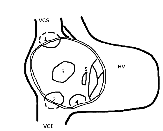 | 568 × 454 (14 KB) | Nja (talk | contribs) | {{Information |Description=Schematic drawing showing the location of different types of ASD, the view is into an opened right atrium. VCS, superior caval vein. VCI, inferior caval vein. HV, right ventricle. 1, upper sinus venosus defect. 2, lower sinus ve |
You cannot overwrite this file.
File usage
The following page uses this file: