File:Figure 8.jpg

Original file (4,606 × 3,224 pixels, file size: 1.27 MB, MIME type: image/jpeg)
| Description |
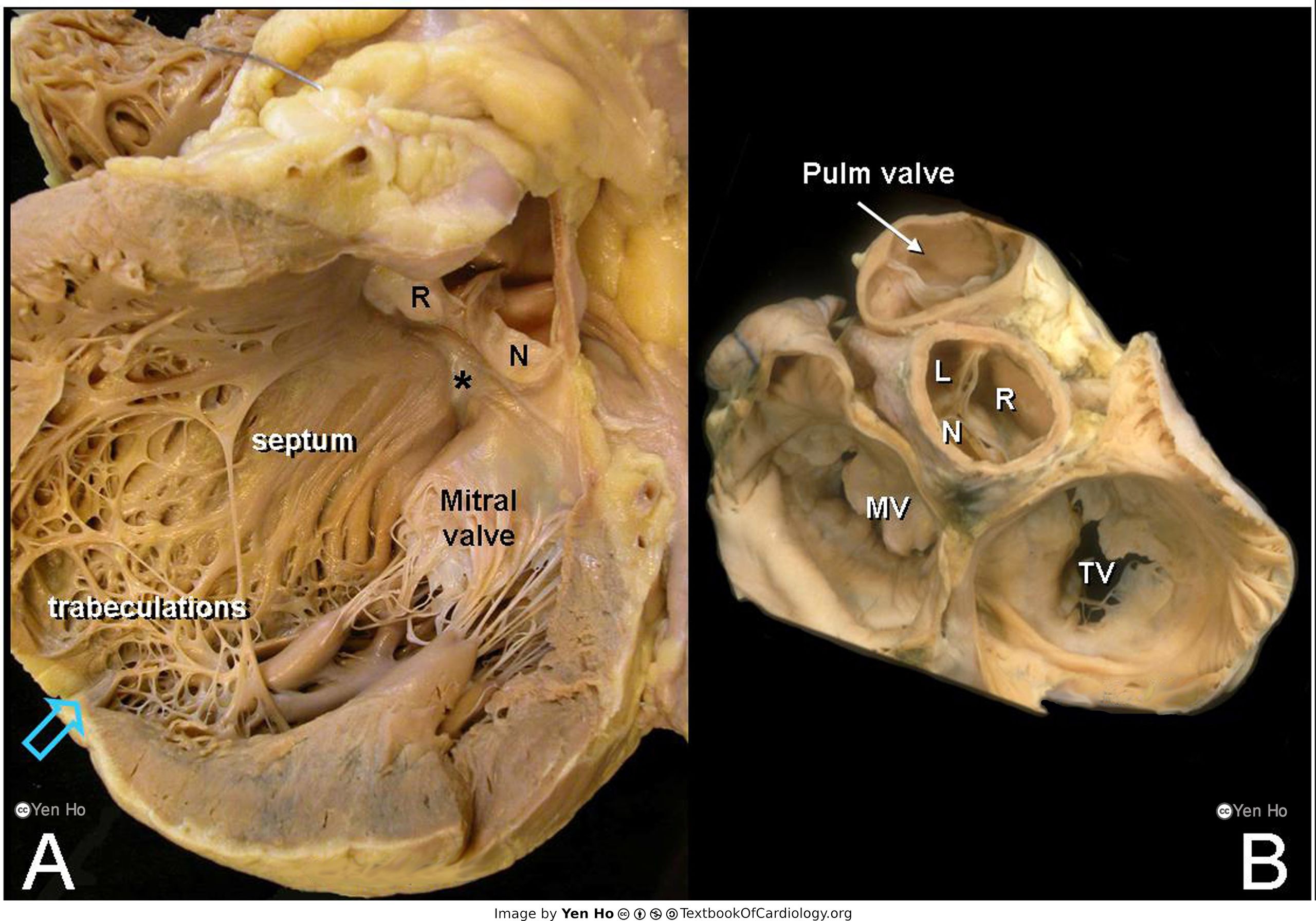
A. The left ventricle is opened through its outflow tract into the aortic valve. The aortic valve leaflets are in fibrous continuity with the anterior leaflet of the mitral valve. The fibrous continuity is expanded at the right and left fibrous trigones. The right trigone(asterisk) is the landmark for the atrioventricular conduction bundle. Note how the thickness of the left ventricular wall diminishes remarkably at the apex (open arrow).
|
|---|---|
| Source |
provided by S. Yen Ho, PhD FRCPath FESC FHEA, Royal Brompton Hospital, UK |
| Date |
2012 |
| Author |
S. Yen Ho, PhD FRCPath FESC FHEA, Royal Brompton Hospital, UK |
| Permission |
File history
Click on a date/time to view the file as it appeared at that time.
| Date/Time | Thumbnail | Dimensions | User | Comment | |
|---|---|---|---|---|---|
| current | 11:10, 18 May 2012 | | 4,606 × 3,224 (1.27 MB) | NiloferT (talk | contribs) |
You cannot overwrite this file.
File usage
The following page uses this file: